Learning Objectives
By the end of this section, you will be able to:
- Describe the structure of eukaryotic plant and animal cells
- State the role of the plasma membrane
- Summarize the functions of the major cellular organelles
- Distinguish cell junctions
At this point, it should be clear that eukaryotic cells have a more complex structure than prokaryotic cells. Organelles allow for various functions to occur in the cell at the same time. Before discussing organelle function, let us first examine two important components of the cell: the plasma membrane and the cytoplasm.
Art Connection

Figure 1: This figure shows (a) a typical animal cell and (b) a typical plant cell.
What structures does a plant cell have that are missing in an animal cell? What structures does an animal cell have that are missing in a plant cell?
The Plasma Membrane
Like prokaryotes, eukaryotic cells have a plasma membrane (Figure 2) made up of a phospholipid bilayer with embedded proteins that separates the internal contents of the cell from its surrounding environment. A more detailed explanation of this structure will be covered in a later chapter. As mentioned in the chemistry unit, a phospholipid is a lipid molecule composed of two fatty acid chains and a phosphate group. The plasma membrane regulates the passage of materials into and out of the cell.

Figure 2. The plasma membrane is a phospholipid bilayer with embedded proteins. There are other components, such as cholesterol and carbohydrates, which can be found in the membrane in addition to phospholipids and protein.
The plasma membranes of cells that specialize in absorption are folded into fingerlike projections called microvilli (singular = microvillus). This folding increases the surface area of the plasma membrane. Such cells are typically found lining the small intestine, the organ that absorbs nutrients from digested food. This is an excellent example of how important structure is to function.
The Cytoplasm
The cytoplasm comprises the contents of a cell between the plasma membrane and the nucleus. It consists of organelles suspended in the gel-like cytosol and the cytoskeleton (Figure 1). The cytoplasm consists of 70-80% water, but has a semi-solid consistency, due to the proteins found within it. Various other organic molecules are found within the cytoplasm allowing for many metabolic reactions, such as protein synthesis, take place.
The Cytoskeleton

Figure 3. Microfilaments, intermediate filaments, and microtubules compose a cell’s cytoskeleton.
If you were to remove all the organelles from a cell, would the plasma membrane and the cytoplasm be the only components that remain? No. Within the cytoplasm, there is a network of protein fibers that has many functions including:
- helps to maintain cell shape;
- secures certain organelles in specific positions;
- allows cytoplasm and vesicles movement within the cell; and
- enables unicellular organisms to move independently.
Collectively, this network of protein fibers is known as the cytoskeleton. There are three types of fibers within the cytoskeleton (Figure 3).
Microfilaments, also know as actin filaments, are the thinnest of the cytoskeletal fibers and function in moving cellular components. This is seen quite a lot during cell division. Common in muscle cells, these components are responsible for muscle cell contraction. Intermediate filaments are important in maintaining the shape of the cell and anchoring organelles. Microtubules are the thickest of the cytoskeletal fibers. These are hollow tubes that can dissolve and reform quickly. Microtubules guide organelle movement and are the structures that pull chromosomes to their poles during cell division. In cilia and flagella, the microtubules are organized as a circle of nine double microtubules on the outside and two microtubules in the center.
The centrosome is a region near the nucleus of animal cells that functions as a microtubule-organizing center. It contains a pair of centrioles, two structures that lie at right angles to each other. The region replicates before a cell divides. The centrioles function by pulling the duplicated chromosomes to opposite ends of a dividing cell. Most plant cells lack centrioles leading to the idea that their role is still not completely understood.
Flagella and Cilia
Flagella (singular = flagellum) are long, hair-like structures extending from the plasma membrane and are used to move an entire cell. When flagella are present, the numbers are fairly small. Cilia are short, hair-like structures that are used to move entire cells or substances along the outer surface of the cell. When cilia(singular = cilium) are present, the numbers are high and extend along the entire surface of the plasma membrane.
The Endomembrane System
The endomembrane system (endo = within) is a group of membranes and organelles (Figure 4) in eukaryotic cells that work together to modify, package, and transport lipids and proteins. It includes the nucleus, the endoplasmic reticulum, and vesicles, the Golgi apparatus, lysosomes, and vacuoles/vesicles. Although not technically within the cell, the plasma membrane is included in the endomembrane system because, as you will see, it interacts with the other endomembranous organelles.
The Nucleus
Typically, the nucleus is the most prominent organelle in a cell (Figure 4). The nucleus (plural = nuclei) houses the cell’s DNA in the form of chromatin and directs the synthesis of ribosomes and proteins. Let us look at it in more detail (Figure 4).

Figure 4. The outermost boundary of the nucleus is the nuclear envelope. Notice that the nuclear envelope consists of two phospholipid bilayers (membranes)—an outer membrane and an inner membrane—in contrast to the plasma membrane, which consists of only one phospholipid bilayer. (credit: modification of work by NIGMS, NIH)
The nuclear envelope is a double-membrane structure that constitutes the outermost portion of the nucleus (Figure 4).
The nuclear envelope is embedded with pores that permit the passage of ions, molecules, and RNA between the nucleus and cytoplasm. The nucleus contains chromatin, thread-like DNA and proteins. Chromatin changes to chromosomes at cell division. In eukaryotes, chromosomes are linear structures. Every species has a specific number of chromosomes in the nucleus of its body cells. For example, in humans, the chromosome number is 46, whereas in fruit flies, the chromosome number is 8.
Chromosomes are only visible and distinguishable from one another just before cell division. When the cell is in the growth and maintenance phases of its life cycle, the chromosomes resemble an unwound, jumbled bunch of threads(chromatin).
The nucleus directs the synthesis of ribosomes, but how? A darkly staining area within the nucleus, called the nucleolus (plural = nucleoli), collecs the ribosomal RNA with associated proteins to assemble the ribosomal subunits. These subunits are then transported through the nuclear pores into the cytoplasm to form ribosomes.
The Endoplasmic Reticulum
The endoplasmic reticulum (ER) (Figure 5) is a series of interconnected membranous tubules, continuous with the nuclear envelope, that collectively modify proteins and synthesize lipids. However, these two functions are performed in separate areas of the endoplasmic reticulum: the rough endoplasmic reticulum and the smooth endoplasmic reticulum, respectively.
The rough endoplasmic reticulum (RER) is so named because the ribosomes attached to its cytoplasmic surface giving it a studded appearance when viewed through an electron microscope. These ribosomes synthesize proteins that are transferred into the lumen of the RER where they undergo modifications. The RER is also responsible for the production of phospholipids making up the plasma membrane. If the phospholipids or modified proteins are not destined to stay in the RER, they will be packaged and transported from the RER by budding from the membrane (Figure 4). The RER is abundant in cells that secrete proteins, such as the liver.
The smooth endoplasmic reticulum (SER) is continuous with the RER but has few or no ribosomes on its cytoplasmic surface (see Figure 4). The functions of SER include:
(1) synthesis of carbohydrates, lipids, and steroid hormones
(2) detoxification of medications and poisons
(3) alcohol metabolism
(4) storage of calcium ions
The Golgi Apparatus

Figure 5. The Golgi apparatus in this transmission electron micrograph of a white blood cell is visible as a stack of semicircular flattened rings in the lower portion of this image. Several vesicles can be seen near the Golgi apparatus. (credit: modification of work by Louisa Howard; scale-bar data from Matt Russell)
Before reaching their final destination, the lipids or proteins for transport need to be sorted, packaged, and tagged so that they arrive at their proper site. The sorting, tagging, packaging, and distribution of lipids and proteins take place in the Golgi apparatus (also called the Golgi body/complex), a series of flattened membranous sacs (Figure 5).
The transport vesicles that form from the ER travel to the Golgi. They fuse with it emptying their contents into the lumen of the Golgi apparatus. As the proteins and lipids travel through the Golgi, they undergo further modifications. The most frequent modification is the addition of short chains of sugar molecules. The newly modified proteins and lipids are tagged for routing to their proper destinations.
Finally, the modified and tagged proteins are packaged into vesicles that bud from the opposite face of the Golgi. Transport vesicles deposit their contents into other parts of the cell for later use. Secretory vesicles fuse with the plasma membrane releasing their contents outside the cell. The number of Golgi in different cell types illustrates the importance of form following function within cells. Cells that engage in a great deal of secretory activity have an abundant number of Golgi. Examples of this can be found in cells of the salivary glands and our immune system where enzymes and antibodies are continuously secreted.
In plant cells, the Golgi has an additional role of synthesizing polysaccharides. Some of them are incorporated into the cell wall and some o are used in other parts of the cell.
Lysosomes
In animal cells, the lysosomes are the cell’s “garbage disposal.” Digestive enzymes within the lysosomes aid the breakdown of proteins, polysaccharides, lipids, nucleic acids, and even worn-out organelles. In single-celled eukaryotes, lysosomes are important for digestion of ingested food and the recycling of organelles. These enzymes are active at a much lower pH (more acidic) than those located in the cytoplasm.
Lysosomes also use their hydrolytic enzymes to destroy disease-causing organisms that might enter the cell. A good example of this occurs as part of our body’s immune system. In a process known as phagocytosis, a section of the plasma membrane of the macrophage(white blood cell) folds in and engulfs a pathogen. The invaginated section containing the pathogen, pinches itself off from the plasma membrane and becomes a vesicle. The vesicle fuses with a lysosome where the hydrolytic enzymes then destroy the pathogen (Figure 6).

Figure 6. A macrophage has phagocytized a potentially pathogenic bacterium into a vesicle, which then fuses with a lysosome within the cell so that the pathogen can be destroyed. Other organelles are present in the cell, but for simplicity, are not shown.
Vesicles and Vacuoles
Vesicles and vacuoles are membrane-bound sacs that function in storage and transport. Vesicles can fuse with other membranes within the cell system while vacuoles do not fuse with other cellular components. Vacuoles are larger than vesicles in all eukaryotic cells. In plants, vacuoles are especially large taking up a great deal of space within the cell.
Art Connection
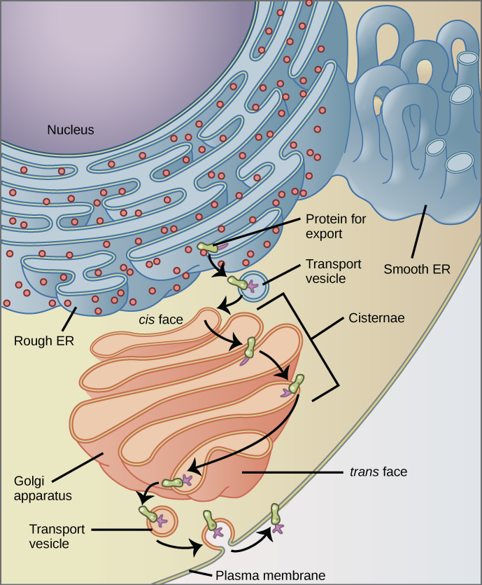
Figure 7. The endomembrane system works to modify, package, and transport lipids and proteins. (credit: modification of work by Magnus Manske)
Why does the cis face of the Golgi not face the plasma membrane?
Ribosomes

Figure 8. Ribosomes are made up of a large subunit (top) and a small subunit (bottom). During protein synthesis, ribosomes assemble amino acids into proteins.
Ribosomes are the cellular structures responsible for protein synthesis. When viewed through an electron microscope, free ribosomes appear as either clusters or single tiny dots floating freely in the cytoplasm. As mentioned earlier, ribosomes can also be attached to the endoplasmic reticulum. Electron microscopy has shown that ribosomes consist of large and small subunits (Figure 8).
Because protein synthesis is essential for all cells, ribosomes are found in practically every cell. In prokaryotic cells, ribosomes generally are smaller. They are particularly abundant in immature red blood cells for the synthesis of hemoglobin, which functions in the transport of oxygen throughout the body.
Mitochondria

Figure 9. This transmission electron micrograph shows a mitochondrion as viewed with an electron microscope. Notice the inner and outer membranes, the cristae, and the mitochondrial matrix. (credit: modification of work by Matthew Britton; scale-bar data from Matt Russell)
Mitochondria (singular = mitochondrion) are often called the “powerhouses” or “energy factories” of a cell because they are responsible for making adenosine triphosphate (ATP), the cell’s main energy-carrying molecule. The formation of ATP from the breakdown of glucose is known as cellular respiration. Mitochondria are oval-shaped, double-membrane organelles (Figure 9) that have their own ribosomes and DNA The inner layer has folds called cristae, which increase the surface area of the inner membrane. The internal area surrounding the folds is known as the matrix. The number of mitochondria vary in a cell. Muscle cells have a very high concentration of mitochondria due to the high energy required for muscle contraction. This is in keeping with form following function.
Peroxisomes
Peroxisomes are small, round organelles enclosed by single membranes. Peroxisomes carry out oxidation reactions that break down fatty acids and amino acids. Alcohol is detoxified by peroxisomes in our liver cells. A byproduct of these reactions is hydrogen peroxide, H2O2, which is contained within the peroxisomes to prevent damage to other cellular components. Hydrogen peroxide is safely broken down by peroxisomal enzymes into water and oxygen.
Animal Cells versus Plant Cells
Despite their fundamental similarities, there are some striking differences between animal and plant cells (see Table). Animal cells have centrioles, centrosomes, and lysosomes, whereas plant cells do not. Plant cells have a cell wall, chloroplasts, plasmodesmata, and plastids and a large central vacuole, whereas animal cells do not.
The Cell Wall
In Figure 1b, the diagram of a plant cell, you see a structure external to the plasma membrane called the cell wall. The cell wall is a rigid covering that protects the cell, provides structural support, and gives shape to the cell. Cell walls are also found in fungal and protist cells.
The major organic molecule in the plant cell wall is cellulose (Figure 10), a polysaccharide made up of long, straight chains of glucose units. When nutritional information refers to dietary fiber, it is referring to the cellulose content of food.

Figure 10. Cellulose is a long chain of β-glucose molecules connected by a 1-4 linkage. The dashed lines at each end of the figure indicate a series of many more glucose units. The size of the page makes it impossible to portray an entire cellulose molecule.
Chloroplasts
Like mitochondria, chloroplasts also have their own DNA and ribosomes. Chloroplasts function in photosynthesis and can be found in eukaryotic cells such as plants and algae. In photosynthesis, carbon dioxide, water, and light energy are used to make glucose and oxygen. Plants (autotrophs) are able to make their own food, like glucose, whereas animals (heterotrophs) must rely on other organisms for their organic compounds or food source.

Figure 11. This simplified diagram of a chloroplast shows the outer membrane, inner membrane, thylakoids, grana, and stroma.
Like mitochondria, chloroplasts have outer and inner membranes, but within the space enclosed by a chloroplast’s inner membrane is a set of interconnected and stacked, fluid-filled membrane sacs called thylakoids (Figure 11). This is the site of photosynthesis. Each stack of thylakoids is called a granum (plural = grana) surrounded by a fluid, stroma.
The chloroplasts contain a green pigment called chlorophyll, which captures the energy of sunlight for photosynthesis. Like plant cells, photosynthetic protists also have chloroplasts. Some bacteria also perform photosynthesis, but they do not have chloroplasts. Their photosynthetic pigments are located in the thylakoid membrane within the cell itself.
Evolution in Action: Endosymbiosis
We have mentioned that both mitochondria and chloroplasts contain DNA and ribosomes. Wonder why? Strong evidence points to endosymbiosis. Endosymbiosis (endo-= within) is a relationship in which one organism lives inside the other. Endosymbiotic relationships abound in nature. An example of this is the microbes living inside the human large intestine. The microbes benefit because they are protected from other organisms and are provided a stable habitat and abundant food supply. We benefit from absorbing the vitamin K produced by the microbes that we would otherwise not obtain.
Scientists have long noticed that bacteria, mitochondria, and chloroplasts are similar in size. We also know that mitochondria and chloroplasts have DNA and ribosomes, just as bacteria do. Scientists believe that host cells and bacteria formed a mutually beneficial endosymbiotic relationship. This occurred when the host cells ingested aerobic bacteria and cyanobacteria but did not destroy them. Through evolution, these ingested bacteria became more specialized in their functions, with the aerobic bacteria becoming mitochondria and the photosynthetic bacteria becoming chloroplasts.
The Central Vacuole
If you look at Figure 1b, you will notice that plant cells each have a large, central vacuole that occupies most of the cell. The central vacuole plays a key role in regulating the water concentration of the cell in changing environmental conditions. In plant cells, the liquid inside the central vacuole provides turgor pressure, which is the outward pressure caused by the fluid inside the cell. What happens if you forget to water your plant? As the water concentration in the soil becomes lower than the water concentration in the plant, water moves out of the central vacuoles and cytoplasm and into the soil. As the central vacuole shrinks, it leaves the cell wall unsupported. This loss of support to the cell walls of a plant results in the wilted appearance.
Extracellular Matrix of Animal Cells

Figure 12. The extracellular matrix consists of a network of substances secreted by cells.
The extracellular matrix is a protective mesh-like network of proteins and polysaccharides. It varies in consistency and works closely with the cytoskeleton to maintain cell shape. (Figure 12). The extracellular matrix hold the cells together to form a tissue and allows the cells within the tissue to communicate with each other.
Intercellular Junctions: Animal vs. Plant
Cells also communicate with each other by direct contact. Intracellular junctions occur in three distinct types within an animal cell:
(1) adhesion junctions(desmosomes) – act like spot welds between adjacent epithelial cells(Figure 13c); keep cells together in sheet-like formation
(2) tight junctions – watertight seal between two adjacent animal cells(Figure 13b); prevents material from leaking between cells; typically found in epithelial tissue lining internal organs/cavities and skin
(3) gap junctions – channels between adjacent cells allowing transport of materials; enable cells to communicate(Figure 13d)
Plasmodesmata (singular = plasmodesma) are junctions between plant cells. Generally in plants, long stretches of the plasma membranes of neighboring plant cells cannot touch one another due to the cell walls surrounding each cell. Plasmodesmata are numerous channels that pass between the cell walls of adjacent plant cells. They connect their cytoplasm and enable signal molecules and nutrients to be transported from cell to cell (Figure 13a).
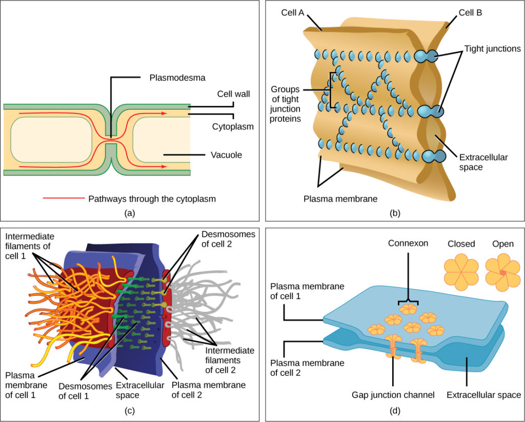
Figure 13. There are four kinds of connections between cells. (a) A plasmodesma is a channel between the cell walls of two adjacent plant cells. (b) Tight junctions join adjacent animal cells. (c) Desmosomes join two animal cells together. (d) Gap junctions act as channels between animal cells. (credit b, c, d: modification of work by Mariana Ruiz Villareal)
COMPARISION OF PROKARYOTIC AND EUKARYOTIC CELLULAR ORGANELLES
| Components of Prokaryotic and Eukaryotic Cells and Their Functions | ||||
|---|---|---|---|---|
| Cell Component | Function | Present in Prokaryotes? | Present in Animal Cells? | Present in Plant Cells? |
| Plasma membrane | Separates cell from external environment; controls passage of organic molecules, ions, water, oxygen, and wastes into and out of the cell | Yes | Yes | Yes |
| Cytoplasm | Provides structure to cell; site of many metabolic reactions; medium in which organelles are found | Yes | Yes | Yes |
| Nucleoid | Location of DNA | Yes | No | No |
| Nucleus | Cell organelle that houses DNA and directs synthesis of ribosomes and proteins | No | Yes | Yes |
| Ribosomes | Protein synthesis | Yes | Yes | Yes |
| Mitochondria | ATP production/cellular respiration | No | Yes | Yes |
| Peroxisomes | Oxidizes and breaks down fatty acids and amino acids, and detoxifies poisons | No | Yes | Yes |
| Vesicles and vacuoles | Storage and transport; digestive function in plant cells | No | Yes | Yes |
| Centrosome | Unspecified role in cell division in animal cells; source of microtubules in animal cells | No | Yes | No |
| Lysosomes | Digestion of macromolecules; recycling of worn-out organelles | No | Yes | No |
| Cell wall | Protection, structural support and maintenance of cell shape | Yes, primarily peptidoglycan in bacteria but not Archaea | No | Yes, primarily cellulose |
| Chloroplasts | Photosynthesis | No | No | Yes |
| Endoplasmic reticulum | Modifies proteins and synthesizes lipids | No | Yes | Yes |
| Golgi apparatus | Modifies, sorts, tags, packages, and distributes lipids and proteins | No | Yes | Yes |
| Cytoskeleton | Maintains cell’s shape, secures organelles in specific positions, allows cytoplasm and vesicles to move within the cell, and enables unicellular organisms to move independently | Yes | Yes | Yes |
| Flagella | Cellular locomotion | Some | Some | No, except for some plant sperm. |
| Cilia | Cellular locomotion, movement of particles along extracellular surface of plasma membrane, and filtration | No | Some | No |
Section Summary
Like a prokaryotic cell, a eukaryotic cell has a plasma membrane, cytoplasm, and ribosomes, but there are a number of differences. A eukaryotic cell is typically larger, has a true nucleus, and has other membrane-bound organelles that allow for compartmentalization of functions. These organelles vary in animal and plant cells but each one provides a function for an overall successful cell life.
Animal cells communicate through their extracellular matrices and are connected to each other by various intracellular junctions. Plant cells are connected and communicate with each other by plasmodesmata, yet another difference between animal and plant cells.
Additional Self Check Questions
1. What structures does a plant cell have that an animal cell does not? What structures does an animal cell have that a plant cell does not?
3. In the context of cell biology, what do we mean by form follows function? What are at least two examples of this concept?
Answers
1. Plant cells have plasmodesmata, a cell wall, a large central vacuole, chloroplasts, and plastids. Animal cells have lysosomes and centrosomes.
2. The plasma membrane interacts with other organelles within the endomembrane system.
3. “Form follows function” refers to the idea that the function of a body part dictates the form of that body part. As an example, organisms like birds or fish that fly or swim quickly through the air or water have streamlined bodies that reduce drag. At the cellular level, in tissues involved in secretory functions, such as the salivary glands, the cells have abundant Golgi.