Spinal Nerves
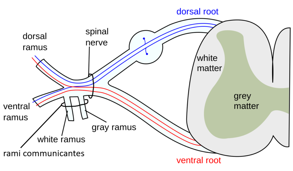
The nerves connected to the spinal cord are the spinal nerves. The arrangement of these nerves is much more regular than that of the cranial nerves. All of the spinal nerves are combined sensory and motor axons that separate into two nerve roots. The sensory axons enter the spinal cord as the dorsal nerve root. The motor fibers, both somatic and autonomic, emerge as the ventral nerve root. The dorsal root ganglion for each nerve is an enlargement of the spinal nerve.
There are 31 spinal nerves, named for the level of the spinal cord at which each one emerges. There are eight pairs of cervical nerves designated C1 to C8, twelve thoracic nerves designated T1 to T12, five pairs of lumbar nerves designated L1 to L5, five pairs of sacral nerves designated S1 to S5, and one pair of coccygeal nerves. The nerves are numbered from the superior to inferior positions, and each emerges from the vertebral column through the intervertebral foramen at its level. The first nerve, C1, emerges between the first cervical vertebra and the occipital bone. The second nerve, C2, emerges between the first and second cervical vertebrae. The same occurs for C3 to C7, but C8 emerges between the seventh cervical vertebra and the first thoracic vertebra. For the thoracic and lumbar nerves, each one emerges between the vertebra that has the same designation and the next vertebra in the column. The sacral nerves emerge from the sacral foramina along the length of that unique vertebra.
Where each spinal nerve exits the vertebrae, it splits and forms branches called rami (singular ramus), shown in Figure 1. The dorsal ramus supplies the posterior of the body with sensory and motor nerves. The rami communicantes is made up of two branches, and carries signals that are part of sympathetic signalling pathways. The ventral ramus provides sensory and motor nerves to the lateral and ventral aspects of the body.
In the thoracic region, the ventral rami of spinal nerves T2-T12 form intercostal nerves

Figure 2. Nerve Plexuses of the Body There are four main nerve plexuses in the human body. The cervical plexus supplies nerves to the posterior head and neck, as well as to the diaphragm. The brachial plexus supplies nerves to the arm. The lumbar plexus supplies nerves to the anterior leg. The sacral plexus supplies nerves to the posterior leg.
that supply the ventral and lateral areas of the chest and superior abdomen. For every other spinal nerve, the axons of the ventral rami combine with those of nearby spinal nerves to form a plexus. In this instance, the word plexus is used to describe networks of nerve fibers with no associated cell bodies. Of the four nerve plexuses, two are found at the cervical level, one at the lumbar level, and one at the sacral level, forming the cervical plexus, brachial plexus, lumbar plexus, and saccral plexus, respectively (Figure 2). Out of each plexus emerge nerves that collect sensory information from, or send motor output to, specific areas or targets in the body. Examples of the areas served by each plexus, and major associated nerves, are given below.
The cervical plexus is composed of axons from spinal nerves C1 through C5 and branches into nerves in the posterior neck and head, where it receives sensory input from the hear neck and superior portion of the shoulder. Motor output is provided to the neck and scapula. Motor signals from this plexus are also sent to the diaphragm muscle at the base of the thoracic cavity via the phrenic nerve.
The other plexus from the cervical level is the brachial plexus. The ventral rami of spinal nerves C5 through T1 reorganize through this plexus to give rise to the nerves of the arms, as the name brachial suggests. The reorganization of axons three times before the major nerves are formed that stem from this plexus, as shown in Figure 3. First, the bundled axons from the ventral rami of C5 to T1 exist separately as roots. Next the axons of C5 and C6 and of C8 and T1 merge, while C7 axons remain separate to form three trunks of the brachial plexus. The posterior Next, axons split off and form six divisions. Finally, axons reorganize to produce three cords: a lateral cord, a posterior cord, and medial cord. Axons from the posterior cord form two nerves: the axillary nerve which provides sensory and motor innervation to the shoulder, and the radial nerve which carries sensory input from the posterior skin of upper limb, forearm, & lateral portion of hand (near the thumb) as well as motor output to the extensor forearm muscles. The medial cord forms the ulnar nerve, which provide motor output to the flexor muscles of forearm and receives sensory input from the medial skin of hand (near the pinky finger). The lateral cord gives rise to the musculocutaneous nerve which carries motor output to the proximal flexor muscles of arm and gets sensory input from lateral forearm. Contributions of axons from both the lateral and medial cords form the median nerve which receives sensory input from the Skin of anterior forearm & hand, and send motor output to the flexor muscles of the forearm.
The lumbar plexus arises from some small contributions from the ventral ramus of T12, and from the ventral remi of L1-L4 spinal nerves. Nerves from this plexus enervate the pelvic region and the anterior leg. Generally, this plexus receives sensory input from the inferior abdomen, pelvic region, legs, and foot – with motor output being sent to the abdominal muscles and muscles of leg. The femoral nerve is one of the major nerves from this plexus, with axon contributions from L2-L4. The femoral gives rise to the saphenous nerve as a branch that extends through the anterior lower leg. Axons of the femoral nerve provide motor control for the quadriceps group, and carries sensory signals for the skin of anterior thigh, and medial leg from knee to foot.
The sacral plexus comes from the lower lumbar nerves L4 and L5 and the sacral nerves S1 to S4. The major areas served by this plexus include the sensory sensations from the pelvic region, leg, and foot – and motor output to the gluteus muscles, leg
muscles, and sphincter muscles of urethra and rectum. The most significant systemic nerve to come from this plexus is the sciatic nerve, formed from axons of L4-S3 making this the largest nerve both in length and number of axons in the body! The sciatic is made from a combination of the axons from the tibial nerve and the fibular nerve, which branch distally at the knee to form two separate nerves. The sciatic nerve sends motor signals to the knee flexors, and gains sensory input from the
skin over posterior thigh. The tibial nerve also sends motor output to the flexors of knee, as well as those used for plantar
flexion of foot & flexion of toes – with sensory signals coming from the skin over posterior leg & sole of foot. Areas served by the fibular nerve include muscles that are flexors of knee, muscles used for dorsiflexion of foot, and extensors of toes – with sensory input from the skin over anterior leg & lateral foot. The sciatic nerve extends across the hip joint and is most commonly associated with the condition sciatica, which is the result of compression or irritation of the nerve or any of the spinal nerves giving rise to it.
The coccygeal plexus is a small plexus served by the axons of the ventral rami from spinal nerves S4, S5, and Co1. It provides sensory innervation to the skin around the area of the coccyx.
Dermatomes
Individual sensory neurons that are in proximity to each other are bundled together into nerves that enter the spinal cord through its posterior root or horn. The region of the skin that each spinal nerve carries information from is called its dermatome. Because there are 31 spinal nerves, there are an equal number of dermatomes, each named for the spinal nerve to which it sends information.

A dermatome map is particularly useful in the diagnosis of spinal cord injuries that interrupt spinal cord transmission. An injured person will lose sensation from all their dermatomes below the level of injury.
Candela Citations
- Authored by: Mysid (original by Tristanb). Located at: https://commons.wikimedia.org/wiki/File:Spinal_nerve.svg. License: CC BY-SA: Attribution-ShareAlike
- Authored by: Open Learning Initiative. Provided by: Carnegie Mellon University. Located at: https://oli.cmu.edu/jcourse/workbook/activity/page?context=4348999280020ca600212ee04a7f1795. Project: Anatomy and Physiology. License: CC BY-NC-SA: Attribution-NonCommercial-ShareAlike
- Anatomy & Physiology. Authored by: OpenStax College . Provided by: Rice University. Located at: http://cnx.org/contents/14fb4ad7-39a1-4eee-ab6e-3ef2482e3e22@9.1. Project: Anatomy & Physiology. License: CC BY: Attribution
- Authored by: Original uploader was Mattopaedia at en.wikipedia derivative work: Captain-n00dle (talk), MissMJ. Located at: https://en.wikipedia.org/wiki/File:Brachial_plexus_2.svg. License: Public Domain: No Known Copyright

