Brain Stem
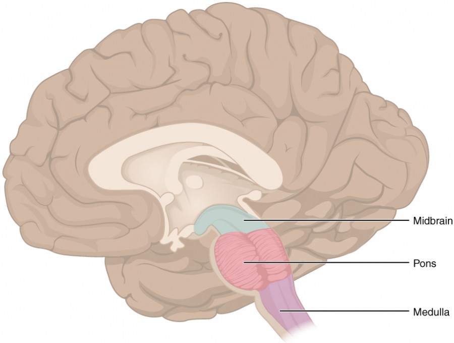
Figure 1. The Brain Stem. The brain stem comprises three regions: the midbrain, the pons, and the medulla.
The midbrain and hindbrain (composed of the pons and the medulla) are collectively referred to as the brain stem (Figure 1). The structure emerges from the ventral surface of the forebrain as a tapering cone that connects the brain to the spinal cord. Attached to the brain stem, but considered a separate region of the adult brain, is the cerebellum. The midbrain coordinates sensory representations of the visual, auditory, and somatosensory perceptual spaces. The pons is the main connection with the cerebellum. The pons and the medulla regulate several crucial functions, including the cardiovascular and respiratory systems and rates.
The cranial nerves connect through the brain stem and provide the brain with the sensory input and motor output associated with the head and neck, including most of the special senses. The major ascending and descending pathways between the spinal cord and brain, specifically the cerebrum, pass through the brain stem.
Medulla Oblongata
The medulla oblongata (or just medulla) is the region known as the myelencephalon in the embryonic brain. The initial portion of the name, “myel,” refers to the significant white matter found in this region—especially on its exterior, which is continuous with the white matter of the spinal cord. The medulla itself is directly attached to the spinal cord, and represents the most inferior area of the brain.

Figure 2. Cross section through medulla.
On the anterior side of the medulla are a pair of large tracts of white matter call the pyramids (Figures 2). The pyramids contain axons of somatic motor neurons from the cerebrum that are transmitting signals through the medulla and towards the spinal cord. At the pyramids, 75-90% of the motor axons decussate, or switch sides so that those that originated on the right cerebral hemisphere move to the left and vice versa. Also visible from the ventral view are a pair of areas of grey matter called the olives (Figures 2). The olives are responsible for relaying sensory information to the cerebellum. Other important areas of grey matter in the medulla include the nucleus cuneatus and nucleus gracilis, both of which are used for relaying sensations to the thalamus to be perceived in the cerebral cortex.

Figure 3. Cross section through the inferior area of the medulla.
A diffuse region of gray matter known as the reticular formation extends from the medulla into the pons and midbrain. It is related to sleep and wakefulness, general brain activity and attention. In brain injuries that damage the reticular formation, individuals fall into a coma as they are unable to generate a state of wakefulness. Within the reticular formation of the medulla are other areas of grey matter that include a cardiovascular center which regulates heart rate and blood pressure, a respiratory center that works in conjunction with the pons to involuntarily regulate breathing rate. Additionally, other centers act to generate involuntary actions in the body such as vomiting, sneezing, hiccuping, and coughing.
Cranial nerves whose nuclei can be found in the medulla include the vestibulocochlear (VIII), glossopharyngeal (CN IX), vagus (CN X), and hypoglossal (XII). (Figure 7)
Pons
The word pons comes from the Latin word for bridge. It is visible on the anterior surface of the brain stem as the thick bundle of white matter. The pons is the main connection between the cerebrum, cerebellum and the brain stem. Large tracts within the pons include the cerebellar peduncles, which carry axons for motor and sensory signals to the the cerebellum. Pyramidal motor tracts in the pons carry somatic motor signals from the cerebrum along a descending path towards the medulla.

Figure 4. Sagittal view of the brainstem showing corticopontine fibers relaying motor signals to the pontine nuclei, which then communicate with the cerebellum via the middle cerebellar peduncle.
The bridge-like white matter is only on the anterior surface of the pons; the gray matter beneath that is a continuation of the tegmentum from the midbrain. Gray matter in the tegmentum region of the pons contains neurons receiving descending input from the cerebral cortex from corticopontine fibers to an area called the pontine nuclei. The pontine nuclei relay motor signals from the cerebrum to the cerebellum via axons in the middle cerebellar peduncle (Figure 4). This allows the cerebellum to modify and correct voluntary muscle movements initiated in the cerebrum. In addition, the reticular formation extends into the pons and forms a respiratory center that works together with that found in the medulla.
Cranial nerves whose nuclei are found in the pons include the trigeminal (CN V), abducens (CN VI), and facial (CN VII). (Figure 7)
Midbrain
One of the original regions of the embryonic brain, the midbrain is a small region between the thalamus and pons. It is separated into the tectum and tegmentum, from the Latin words for roof and floor, respectively. The cerebral aqueduct passes through the center of the midbrain, such that these regions are the roof and floor of that canal.
The tectum is composed of four bumps known as the corpora quadrigemina. Each pair of bumps are called colliculi (singular = colliculus), which means “little hill” in Latin. The inferior colliculi are the inferior pair of these enlargements and is part of the auditory brain stem pathway (Figure 5). Neurons of the inferior colliculi project to the thalamus, which then sends auditory information to the cerebrum for the conscious perception of sound. The inferior colliculi also process sudden sounds to produce a reflex reaction by which the head and eyes involuntarily turn towards the sound. If you were sitting quietly taking a test and heard the person next to you drop their pencil, the sudden sound would cause your inferior colliculi to turn your head and eyes towards the pencil where it sat on the floor. The superior colliculi are the superior pair and combine sensory information about visual space (Figure 5). Activity in the superior colliculi is related to orienting the eyes to track a moving object, to scan an area when looking for something, or to track words on a page while reading. If you were searching a crowded room to locate your friend, the pattern with which your eyes scanned the room would be controlled by the superior colliculi.

Figure 5. Posterior view of brainstem and inferior diencephalon.
The tegmentum is continuous with the gray matter of the rest of the brain stem. Throughout the midbrain, pons, and medulla, the tegmentum contains the nuclei that receive and send information through the cranial nerves, as well as regions that regulate important functions such as those of the cardiovascular and respiratory systems.
Important areas of grey matter on the interior of the midbrain include the red nucleus and the substantia nigra (Figure 6). The red nucleus is a region that possesses a reddish color due to the abundance of blood vessels that flow through this area. Functionally, the red nucleus plays a role in producing involuntary skeletal muscle contractions. The substantia nigra is also visually distinctive in that it possesses neurons with melanin, giving this group of neurons a darker appearance. The neurons of the substantia nigra produce dopamine, which is transported via their axons to the basal nuclei of the cerebrum where the dopamine is used to regulate motor signals.

Figure 6. Cross section through the midbrain showing the superior colliculus, red nucleus, substantia nigra, and cerebral aquaduct.
Areas of white matter include the cerebral peduncles, which can be seen on the anterior view of the midbrain, and the cerebellar peduncles. The cerebral peduncles transmit motor signals along axons from the cerebrum to the midbrain. The cerebellar peduncles allow for communication of sensory and motor signals from the cerebellum to the midbrain and other parts of the brainstem. (Figure 7)
Cranial nerve nuclei found in the midbrain include those for the oculomotor nerve (CN III) and the trochlear nerve (CN IV). (Figure 7)

Figure 7. Anterior view of the brainstem and inferior portion of diencephalon.
Candela Citations
- Anatomy & Physiology. Authored by: Open Stax College. Provided by: Rice University. Located at: http://cnx.org/contents/14fb4ad7-39a1-4eee-ab6e-3ef2482e3e22@8.25. License: CC BY: Attribution. License Terms: Download for free at http://cnx.org/contents/14fb4ad7-39a1-4eee-ab6e-3ef2482e3e22@8.25
- Figures 2-7. Edits by C. Rounds. Authored by: D.J. Cunningham. Provided by: Internet Archive. Located at: https://archive.org/details/cunninghamstextb00cunn. License: Public Domain: No Known Copyright