The nervous system is critical to many of our homeostatic feedback loops. In most of these loops, the structures of the nervous system make up more than one component, and carry out more than one function in these loops. For example, specialized nerve endings often act as sensors (receptors), information is carried along nerves and/or tracts of the spinal cord, integration occurs within the CNS, and spinal cord tracts and nerves carry the responding information back out to the effectors. The spinal cord is a nervous system structure dedicated to relaying information from the periphery to the brain and back, as well as carrying out certain levels of integration, such as those found in many reflexes. The structure of the spinal cord aids it in carrying out these relaying and integrative functions.
Gross Anatomy of the Spinal Cord
Whereas the brain develops out of expansions of the neural tube into primary and then secondary vesicles, the spinal cord maintains the tube structure and is only specialized into certain regions. As the spinal cord continues to develop in the newborn, anatomical features mark its surface. The anterior midline is marked by the anterior median fissure, and the posterior midline is marked by the posterior median sulcus. Axons enter the posterior side through the dorsal (posterior) nerve root, which marks the posterolateral sulcus on either side. The axons emerging from the anterior side do so through the ventral (anterior) nerve root. Note that it is common to see the terms dorsal (dorsal = “back”) and ventral (ventral = “belly”) used interchangeably with posterior and anterior, particularly in reference to nerves and the structures of the spinal cord. You should learn to be comfortable with both.

Figure 1. Inferior spinal cord showing the conus meullaris (medullary cone) and cauda equina.
On the whole, the posterior regions are responsible for sensory functions and the anterior regions are associated with motor
functions. This comes from the initial development of the spinal cord, which is divided into the basal plate and the alar plate. The basal plate is closest to the ventral midline of the neural tube, which will become the anterior face of the spinal cord and gives rise to motor neurons. The alar plate is on the dorsal side of the neural tube and gives rise to neurons that will receive sensory input from the periphery. The length of the spinal cord is divided into regions that correspond to the regions of the vertebral column. The name of a spinal cord region corresponds to the level at which spinal nerves pass through the intervertebral foramina.
Starting between the base of the skull and the first cervical vertebrae, and continuing into the sacral region of the spinal column, a pair of spinal nerves extend from the spinal cord (although information is transmitted in both directions on sensory and motor neurons within these mixed nerves). The nerves that emerge from the spinal cord pass through the intervertebral formina at the respective levels. All but the first spinal nerve (C1) pass through the intervertebral foramen of the spinal cord, whereas spinal nerve C1 passes between the occipital bone and vertebrae C1. In all there are 31 pairs of spinal nerves that carry information to and from the spinal cord and the periphery of the body. Note that not all of the spinal nerves arise from the cord at the level of the vertebrae between which they pass. This is most obvious when considering those spinal nerves arising in the lower lumbar and sacral regions, which are areas of the vertebral column that do not contain any spinal cord. The reason for this is that the spinal cord is not the full length of the vertebral column because the spinal cord does not grow significantly longer after the first or second year, but the skeleton continues to grow. The result is that the end of the spinal cord is located at the conus medullaris (or medullary cone) between L1 and L2. As the vertebral column grows, the spinal nerves grow with it and

Figure 2. Ventral and dorsal views of the spinal cord, with cervical and lumbar enlargements shown.
result in a long bundle of nerves that extend past the conus medullaris which resembles a horse’s tail and is named the cauda equina (Figure 1). Because the spinal nerve roots don’t always originate at the level of the vertebrae that they pass through, the segments of the spinal cord are named for
the spinal nerve to which they give rise. For example, segment S2 of the spinal cord would be located near the T12 vertebrae.
The spinal cord terminates near vertebrae L1, but there is a lot of body tissue that needs to be innervated below this level, so there are a significant number of nerves arising from the lower aspect of the spinal cord. This leads to an area of increased spinal cord thickness in the lumbosacral regions of the spinal cord (corresponding to a region associated with the inferior thoracic vertebrae) called the lumbar enlargement (Figure 2). There is a corresponding cervical enlargement in the cervical segments that give rise to nerves innervating the upper limbs.
Spinal Cord Structure
Recall that the central nervous system tissues can generally be divided into white matter and gray matter. White matter is the myelin-containing region composed of axons, which make up the tracts of the CNS. These carry information between different regions and structures in the CNS. Gray matter contains the cell bodies and dendrites and therefore is the site of synaptic transmission.
Gray Horns
In cross-section, the gray matter of the spinal cord has the appearance of an ink-blot test, with the spread of the gray matter on one side replicated on the other—a shape reminiscent of a bulbous capital “H.” As shown in Figure 3, the gray matter is subdivided into regions that are referred to as horns. The dorsal horn is responsible for sensory processing, and contain interneurons that receive somatic and visceral sensory signals. The ventral horn sends out motor signals to the skeletal muscles. The lateral horn, which is only found in the thoracic, upper lumbar, and sacral regions, sends out signals to visceral targets as part of the autonomic nervous system. The matched horns on each side of the “butterfly” are connected via the gray commissure, which also surrounds the cerebrospinal fluid filled central canal.
Some of the largest neurons of the spinal cord are the multipolar motor neurons in the ventral horn. The fibers that cause contraction of skeletal muscles are the axons of these neurons. The motor neuron that causes contraction of the big toe, for example, is located in the sacral spinal cord. The axon that has to reach all the way to the belly of that muscle may be a meter (about 3 feet) in length. The neuronal cell body that maintains that long fiber must be quite large, possibly several hundred micrometers in diameter, making it one of the largest cells in the body.
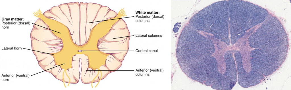
Figure 9. Cross-section of Spinal Cord. The cross-section of a thoracic spinal cord segment shows the posterior, anterior, and lateral horns of gray matter, as well as the posterior, anterior, and lateral columns of white matter. LM × 40. (Micrograph provided by the Regents of University of Michigan Medical School © 2012)
White Columns
Just as the gray matter is separated into horns, the white matter of the spinal cord is separated into columns. Ascending tracts of nervous system fibers in these columns carry sensory information up to the brain, whereas descending tracts carry motor commands from the brain. Looking at the spinal cord longitudinally, the columns extend along its length as continuous bands of white matter. Between the two posterior horns of gray matter are the posterior columns. Between the two anterior horns, and bounded by the axons of motor neurons emerging from that gray matter area, are the anterior columns. The white matter on either side of the spinal cord, between the posterior horn and the axons of the anterior horn neurons, are the lateral columns. The posterior columns are composed of axons of ascending tracts. The anterior and lateral columns are composed of many different groups of axons of both ascending and descending tracts—the latter carrying motor commands down from the brain to the spinal cord to control output to the periphery.
Spinal Nerves
Each segment of the spinal cord possesses a pair of spinal nerves, which possess a mixture of motor and sensory axons. Where the spinal nerve attaches to the spinal cord it splits into two roots. The dorsal root contains only sensory axons. This root is easy to recognize because the unipolar sensory neurons in the root have all of their cell bodies clustered together in one spot which forms a visible bump on the dorsal root called the dorsal root ganglion. In the ventral root there are only the axons of motor neurons, whose cell bodies are located in the ventral or lateral horns of gray matter. As a result, there is no bump on the ventral root.

Figure 3. Transverse section of spinal cord showing spinal nerve root and grey matter components.

Figure 4. Transverse sections through the spinal cord at different levels.
Although each segment of the spinal cord has similar features, there are some differences along its length, as you may be able to determine from the image above. The main difference is that the ratio of gray matter to white matter varies among segments of the spinal cord. At the lower levels of the spinal cord there is a greater ratio of grey matter to white matter. This should make sense, as there are less ascending and descending tracts of whiter matter as you move lower. As previously mentioned, the lateral horns are only found in the thoracic and lumber regions of the spinal cord, where they contain the motor nuclei of the sympathetic nervous system. Finally, the size of the anterior and posterior horns varies, depending on the amount of tissue they are innervating. For example, the thoracic segments have relatively small anterior horns, as there is little skeletal muscle to innervate in the thorax and abdomen, while the cervical and thoracolumbar regions have large anterior horns, used to innervate the skeletal muscles of the arms and legs, respectively.
Watch this video to learn about the gray matter of the spinal cord that receives input from fibers of the dorsal (posterior) root and sends information out through the fibers of the ventral (anterior) root.
As discussed in this video, these connections represent the interactions of the CNS with peripheral structures for both sensory and motor functions. The cervical and lumbar spinal cords have enlargements as a result of larger populations of neurons. What are these enlargements responsible for?
Spinal Cord Tracts
The white matter of the spinal cord is divided into the paired posterior (dorsal), lateral, and anterior (ventral) columns. These columns are sometimes called funiculi (or funiculus when singular) and are made up of axons that are traveling up (ascending) or down (descending) the spinal cord. The ascending tracts generally carry sensory information from the periphery to the brain, while the descending tracts carry motor signals to muscles and glands.
The columns can be further divided into tracts (sometimes called fasciculi), which is a way of functionally grouping the neurons based on similar origin, destination and function. These tracts are often named for the structures that they connect. For example, the spinothalamic tract indicates that the fibers are carrying information from the spinal cord to the thalamus of the brainstem. You may note from its name that it is an ascending tract, so the information that it carries is sensory.
Some of the tracts cross over (decussate) either in the spinal cord or brainstem, and when this occurs, the relationship between the origin and destination is termed contralateral. Much of our motor control is contralateral. For example, your right arm is mainly controlled by the motor area in your left brain. When the origin and destination of a tract are on the same side of the body, it is referred to as an ispsilateral relationship.

Figure 5. Descending and Ascending pathways through the spinal cord.
This table lists the major spinal tracts, indicates if they decussate, and provides a brief description of the types of information that they carry.

Candela Citations
- Unit 14: Nervous System (Module 54). Authored by: Open Learning Initiative. Provided by: Carnegie Mellon University. Located at: https://oli.cmu.edu/jcourse/workbook/activity/page?context=434898bf80020ca6003325b6ff7c29f8. Project: Anatomy & Physiology. License: CC BY-NC-SA: Attribution-NonCommercial-ShareAlike
- Authored by: Open Stax College. Provided by: Rice University. Located at: http://cnx.org/contents/14fb4ad7-39a1-4eee-ab6e-3ef2482e3e22@8.25. License: CC BY: Attribution. License Terms: Download for free at http://cnx.org/contents/14fb4ad7-39a1-4eee-ab6e-3ef2482e3e22@8.25
- Authored by: Polarlys and Mikael Hu00e4ggstru00f6m. Located at: https://commons.wikimedia.org/wiki/File:Spinal_cord_tracts_-_English.svg. License: CC BY-SA: Attribution-ShareAlike
- Authored by: Henry Gray. Located at: https://en.wikipedia.org/wiki/Spinal_cord#/media/File:Gray666.png. License: Public Domain: No Known Copyright