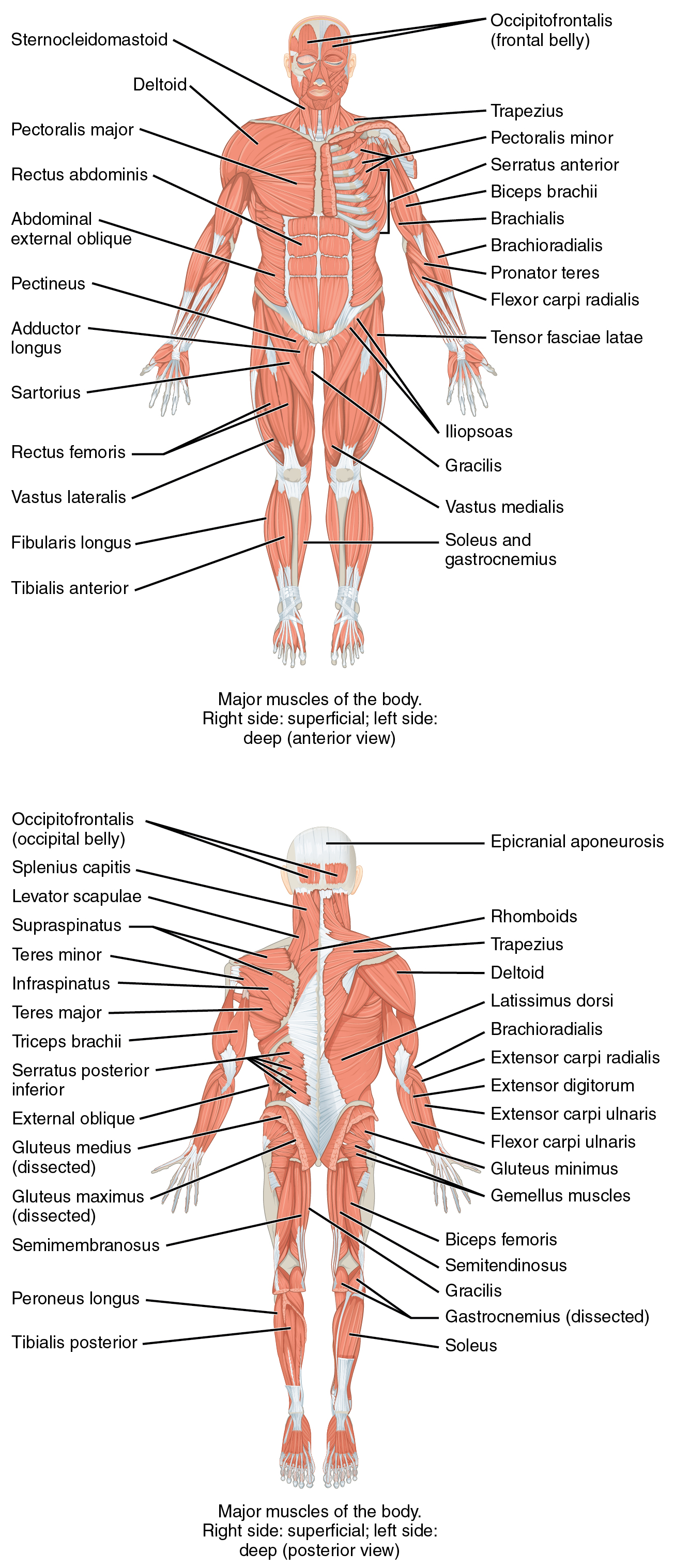
To move the skeleton, the tension created by the contraction of the fibers in most skeletal muscles is transferred to the tendons. The tendons are strong bands of dense, regular connective tissue that connect muscles to bones. The bone connection is why this muscle tissue is called skeletal muscle.Interactions of Skeletal Muscles in the Body
To pull on a bone, that is, to change the angle at its synovial joint, which essentially moves the skeleton, a skeletal muscle must also be attached to a fixed part of the skeleton. The moveable end of the muscle that attaches to the bone being pulled is called the muscle’s insertion, and the end of the muscle attached to a fixed (stabilized) bone is called the origin. During forearm flexion—bending the elbow—the brachioradialis assists the brachialis.

Figure 1. Prime Movers and Synergists. The biceps brachii flex the lower arm. The brachoradialis, in the forearm, and brachialis, located deep to the biceps in the upper arm, are both synergists that aid in this motion.
Although a number of muscles may be involved in an action, the principal muscle involved is called the prime mover, or agonist. To lift a cup, a muscle called the biceps brachii is actually the prime mover; however, because it can be assisted by the brachialis, the brachialis is called a synergist in this action (Figure 1). A synergist can also be a fixator that stabilizes the bone that is the attachment for the prime mover’s origin.
A muscle with the opposite action of the prime mover is called an antagonist. Antagonists play two important roles in muscle function:
- They maintain body or limb position, such as holding the arm out or standing erect
- They control rapid movement, as in shadow boxing without landing a punch or the ability to check the motion of a limb
For example, to extend the knee, a group of four muscles called the quadriceps femoris in the anterior compartment of the thigh are activated (and would be called the agonists of knee extension). However, to flex the knee joint, an opposite or antagonistic set of muscles called the hamstrings is activated.
As you can see, these terms would also be reversed for the opposing action. If you consider the first action as the knee bending, the hamstrings would be called the agonists and the quadriceps femoris would then be called the antagonists. See Table 1 for a list of some agonists and antagonists.
There are also skeletal muscles that do not pull against the skeleton for movements. For example, there are the muscles that produce facial expressions. The insertions and origins of facial muscles are in the skin, so that certain individual muscles contract to form a smile or frown, form sounds or words, and raise the eyebrows. There also are skeletal muscles in the tongue, and the external urinary and anal sphincters that allow for voluntary regulation of urination and defecation, respectively. In addition, the diaphragm contracts and relaxes to change the volume of the pleural cavities but it does not move the skeleton to do this.
Table 1: Major Muscles of the Human Body and their Actions.
Muscles |
Actions |
|
Major Muscles ofUpper Limb |
Deltoid | shoulder abduction, flexion and extension |
| Biceps brachii | flexes elbow | |
| Triceps brachii | extends forearm | |
Major Muscles ofLower Limb |
Gluteus maximus | external rotation and extension of the hip joint, |
| Sartorius | lateral rotation and abduction of thigh; flexion and medial rotation of leg | |
| Quadriceps group (4 muscles together) |
knee extension; hip flexion | |
| Hamstrings group (3 muscles together) |
flexes knee, extends hip joint, medially rotates leg at knee |
|
| Tibialis anterior | dorsiflex and invert the foot | |
| Gastrocnemius | plantar flexes foot – extension or flexion of the foot at the ankle, flexes knee | |
Major Muscles ofTrunk – Anterior |
Pectoralis | control the movement of the arm, create lateral, vertical, or rotational motion. |
| Rectus abdominus | pulls the ribs and the pelvis in and curves the back. (doing crunches ) | |
| External oblique | rotate the trunk | |
Major Muscles ofTrunk – Posterior |
Trapezius (upper back) | tilt and turn the head and neck, shrug, steady the shoulders, and twist the arms. Elevates, depresses, rotates, and retracts the scapula, or shoulder blade |
| Latissimus dorsi (lower back) | When flexed, the muscle works at extending, adducting and rotating the arm. | |
| Table 2. Agonist and Antagonist Skeletal Muscle Pairs | ||
| Agonist | Antagonist | Movement |
|---|---|---|
| Biceps brachii: in the anterior compartment of the arm | Triceps brachii: in the posterior compartment of the arm | The biceps brachii flexes the forearm, whereas the triceps brachii extends it. |
| Hamstrings: group of three muscles in the posterior compartment of the thigh | Quadriceps femoris: group of four muscles in the anterior compartment of the thigh | The hamstrings flex the leg, whereas the quadriceps femoris extend it. |
Everyday Connections: Exercise and Stretching
When exercising, it is important to first warm up the muscles. Stretching pulls on the muscle fibers and it also results in an increased blood flow to the muscles being worked. Without a proper warm-up, it is possible that you may either damage some of the muscle fibers or pull a tendon. A pulled tendon, regardless of location, results in pain, swelling, and diminished function; if it is moderate to severe, the injury could immobilize you for an extended period.
Recall the discussion about muscles crossing joints to create movement. Most of the joints you use during exercise are synovial joints, which have synovial fluid in the joint space between two bones. Exercise and stretching may also have a beneficial effect on synovial joints. Synovial fluid is a thin, but viscous film with the consistency of egg whites. When you first get up and start moving, your joints feel stiff for a number of reasons. After proper stretching and warm-up, the synovial fluid may become less viscous, allowing for better joint function.

Candela Citations
- Chapter 11: The Muscular System. Authored by: OpenStax College. Provided by: Rice University. Located at: http://cnx.org/contents/14fb4ad7-39a1-4eee-ab6e-3ef2482e3e22@7.1@7.1.. Project: Anatomy & Physiology. License: CC BY: Attribution. License Terms: Download for free at http://cnx.org/content/col11496/latest/.