Now that you have learned that the cell membrane surrounds all cells, you can dive inside of a prototypical human cell to learn about its internal components and their functions. All living cells in multicellular organisms contain an internal cytoplasmic compartment, and a nucleus within the cytoplasm. Cytosol, the jelly-like substance within the cell, provides the fluid medium necessary for biochemical reactions. Eukaryotic cells, including all animal cells, also contain various cellular organelles. An organelle (“little organ”) is one of several different types of membrane-enclosed bodies in the cell, each performing a unique function. Just as the various bodily organs work together in harmony to perform all of a human’s functions, the many different cellular organelles work together to keep the cell healthy and performing all of its important functions. The organelles and cytosol, taken together, compose the cell’s cytoplasm. The nucleus is a cell’s central organelle, which contains the cell’s DNA (Figure 1).
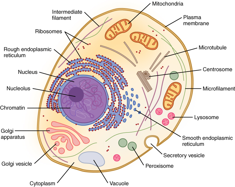
Figure 1. Prototypical Human Cell. While this image is not indicative of any one particular human cell, it is a prototypical example of a cell containing the primary organelles and internal structures.
The Nucleus
Typically, the nucleus is the most prominent organelle in a cell ((Figure 2)). The nucleus (plural = nuclei) houses the cell’s DNA and directs the synthesis of ribosomes and proteins. Let’s look at it in more detail.

Figure 2. The nucleus stores chromatin (DNA plus proteins) in a gel-like substance called the nucleoplasm. The nucleolus is a condensed chromatin region where ribosome synthesis occurs. We call the nucleus’ boundary the nuclear envelope. It consists of two phospholipid bilayers: an outer and an inner membrane. The nuclear membrane is continuous with the endoplasmic reticulum. Nuclear pores allow substances to enter and exit the nucleus.
The Nuclear Envelope
The nuclear envelope is a double-membrane structure that constitutes the nucleus’ outermost portion ((Figure)). Both the nuclear envelope’s inner and outer membranes are phospholipid bilayers. The nuclear envelope is punctuated with pores that control the passage of ions, molecules, and RNA between the nucleoplasm and cytoplasm. The nucleoplasm is the semi-solid fluid inside the nucleus, where we find the chromatin and the nucleolus.
Chromatin and Chromosomes
To understand chromatin, it is helpful to first explore chromosomes, structures within the nucleus that are made up of DNA, the hereditary material. You may remember that in prokaryotes, DNA is organized into a single circular chromosome. In eukaryotes, chromosomes are linear structures. Every eukaryotic species has a specific number of chromosomes in the nucleus of each cell. For example, in humans, the chromosome number is 46, while in fruit flies, it is eight.
Chromosomes are only visible and distinguishable from one another when the cell is getting ready to divide. When the cell is in the growth and maintenance phases of its life cycle, proteins attach to chromosomes, and they resemble an unwound, jumbled bunch of threads. We call these unwound protein-chromosome complexes chromatin ((Figure 3)). Chromatin describes the material that makes up the chromosomes both when condensed and decondensed.


Figure 3. (a) This image shows various levels of chromatin’s organization (DNA and protein). (b) This image shows paired chromosomes. (credit b: modification of work by NIH; scale-bar data from Matt Russell)
The Nucleolus
We already know that the nucleus directs the synthesis of ribosomes, but how does it do this? Some chromosomes have sections of DNA that encode ribosomal RNA. A darkly staining area within the nucleus called the nucleolus (plural = nucleoli) aggregates the ribosomal RNA with associated proteins to assemble the ribosomal subunits that are then transported out through the pores in the nuclear envelope to the cytoplasm.
Organelles of the Endomembrane System
A set of three major organelles together form a system within the cell called the endomembrane system. These organelles work together to perform various cellular jobs, including the task of producing, packaging, and exporting certain cellular products. The organelles of the endomembrane system include the endoplasmic reticulum, Golgi apparatus, and vesicles.
Endoplasmic Reticulum
The endoplasmic reticulum (ER) is a system of channels that is continuous with the nuclear membrane (or “envelope”) covering the nucleus and composed of the same lipid bilayer material. The ER can be thought of as a series of winding thoroughfares similar to the waterway canals in Venice. The ER provides passages throughout much of the cell that function in transporting, synthesizing, and storing materials. The winding structure of the ER results in a large membranous surface area that supports its many functions (Figure 4).

Figure 4. Endoplasmic Reticulum (ER). Click for a larger image. (a) The ER is a winding network of thin membranous sacs found in close association with the cell nucleus. The smooth and rough endoplasmic reticula are very different in appearance and function (source: mouse tissue). (b) Rough ER is studded with numerous ribosomes, which are sites of protein synthesis (source: mouse tissue). EM × 110,000. (c) Smooth ER synthesizes phospholipids, steroid hormones, regulates the concentration of cellular Ca++,metabolizes some carbohydrates, and breaks down certain toxins (source: mouse tissue). EM × 110,510. (Micrographs provided by the Regents of University of Michigan Medical School © 2012)
Endoplasmic reticulum can exist in two forms: rough ER and smooth ER. These two types of ER perform some very different functions and can be found in very different amounts depending on the type of cell. Rough ER (RER) is so-called because its membrane is dotted with embedded granules—organelles called ribosomes, giving the RER a bumpy appearance. A ribosome is an organelle that serves as the site of protein synthesis. It is composed of two ribosomal RNA subunits that wrap around mRNA to start the process of translation, followed by protein synthesis. Smooth ER (SER) lacks these ribosomes. One of the main functions of the smooth ER is in the synthesis of lipids. The smooth ER synthesizes phospholipids, the main component of biological membranes, as well as steroid hormones. For this reason, cells that produce large quantities of such hormones, such as those of the female ovaries and male testes, contain large amounts of smooth ER. In addition to lipid synthesis, the smooth ER also sequesters (i.e., stores) and regulates the concentration of cellular Ca++, a function extremely important in cells of the nervous system where Ca++ is the trigger for neurotransmitter release. The smooth ER additionally metabolizes some carbohydrates and performs a detoxification role, breaking down certain toxins. In contrast with the smooth ER, the primary job of the rough ER is the synthesis and modification of proteins destined for the cell membrane or for export from the cell. For this protein synthesis, many ribosomes attach to the ER (giving it the studded appearance of rough ER). Typically, a protein is synthesized within the ribosome and released inside the channel of the rough ER, where sugars can be added to it (by a process called glycosylation) before it is transported within a vesicle to the next stage in the packaging and shipping process: the Golgi apparatus.
Career Connection
Cardiologist
Heart disease is the leading cause of death in the United States. This is primarily due to our sedentary lifestyle and our high trans-fat diets.
Heart failure is just one of many disabling heart conditions. Heart failure does not mean that the heart has stopped working. Rather, it means that the heart can’t pump with sufficient force to transport oxygenated blood to all the vital organs. Left untreated, heart failure can lead to kidney failure and other organ failure.
Cardiac muscle tissue comprises the heart’s wall. Heart failure occurs when cardiac muscle cells’ endoplasmic reticula do not function properly. As a result, an insufficient number of calcium ions are available to trigger a sufficient contractile force.
Cardiologists (cardi- = “heart”; -ologist = “one who studies”) are doctors who specialize in treating heart diseases, including heart failure. Cardiologists can diagnose heart failure via a physical examination, results from an electrocardiogram (ECG, a test that measures the heart’s electrical activity), a chest X-ray to see whether the heart is enlarged, and other tests. If the cardiologist diagnoses heart failure, he or she will typically prescribe appropriate medications and recommend a reduced table salt intake and a supervised exercise program.
The Golgi Apparatus
The Golgi apparatus is responsible for sorting, modifying, and shipping off the products that come from the rough ER, much like a post-office. The Golgi apparatus looks like stacked flattened discs, almost like stacks of oddly shaped pancakes. Like the ER, these discs are membranous. The Golgi apparatus has two distinct sides, each with a different role. One side of the apparatus receives products in vesicles. These products are sorted through the apparatus, and then they are released from the opposite side after being repackaged into new vesicles. If the product is to be exported from the cell, the vesicle migrates to the cell surface and fuses to the cell membrane, and the cargo is secreted (Figure 5).
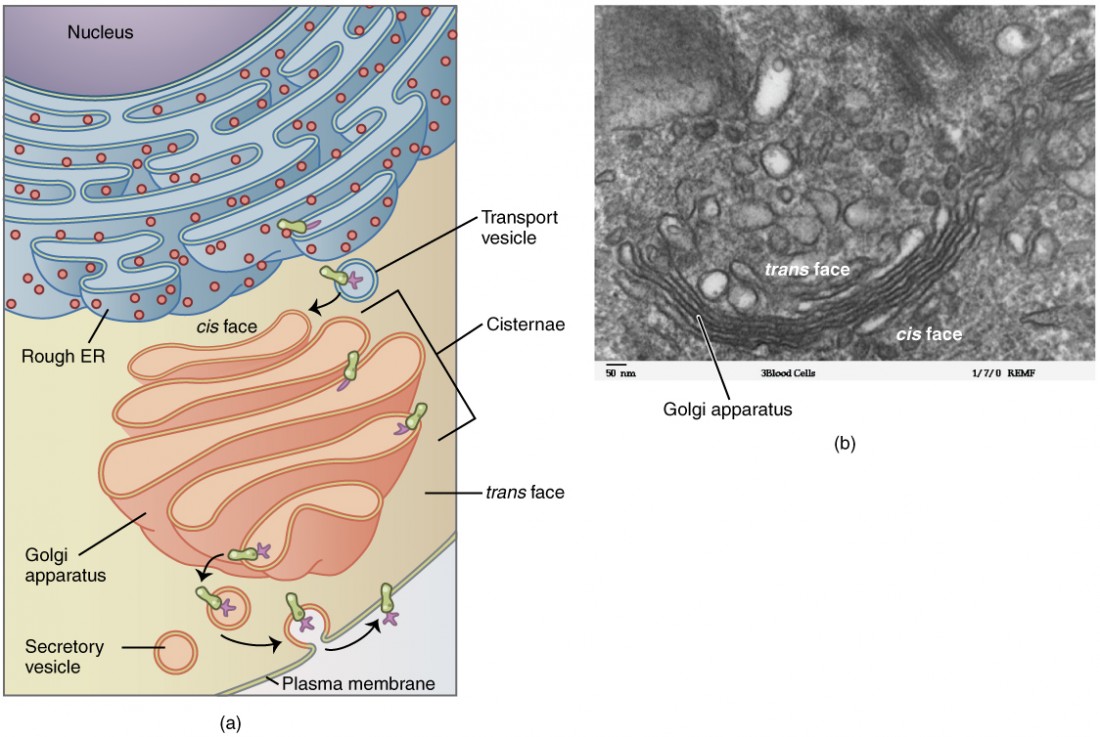
Figure 5. Golgi Apparatus. (a) The Golgi apparatus manipulates products from the rough ER, and also produces new organelles called lysosomes. Proteins and other products of the ER are sent to the Golgi apparatus, which organizes, modifies, packages, and tags them. Some of these products are transported to other areas of the cell and some are exported from the cell through exocytosis. Enzymatic proteins are packaged as new lysosomes (or packaged and sent for fusion with existing lysosomes). (b) An electron micrograph of the Golgi apparatus.
Career Connection
Geneticist
Many diseases arise from genetic mutations that prevent synthesizing critical proteins. One such disease is Lowe disease (or oculocerebrorenal syndrome, because it affects the eyes, brain, and kidneys). In Lowe disease, there is a deficiency in an enzyme localized to the Golgi apparatus. Children with Lowe disease are born with cataracts, typically develop kidney disease after the first year of life, and may have impaired mental abilities.
A mutation on the X chromosome causes Lowe disease. The X chromosome is one of the two human sex chromosomes, as these chromosomes determine a person’s sex. Females possess two X chromosomes while males possess one X and one Y chromosome. In females, the genes on only one of the two X chromosomes are expressed. Females who carry the Lowe disease gene on one of their X chromosomes are carriers and do not show symptoms of the disease. However, males only have one X chromosome and the genes on this chromosome are always expressed. Therefore, males will always have Lowe disease if their X chromosome carries the Lowe disease gene. Geneticists have identified the mutated gene’s location, as well as many other mutation locations that cause genetic diseases. Through prenatal testing, a woman can find out if the fetus she is carrying may be afflicted with one of several genetic diseases.
Geneticists analyze prenatal genetic test results and may counsel pregnant women on available options. They may also conduct genetic research that leads to new drugs or foods, or perform DNA analyses for forensic investigations.
Lysosomes
Some of the protein products packaged by the Golgi include digestive enzymes that are meant to remain inside the cell for use in breaking down certain materials. The enzyme-containing vesicles released by the Golgi may form new lysosomes, or fuse with existing, lysosomes. A lysosome is an organelle that contains enzymes that break down and digest unneeded cellular components, such as a damaged organelle. (A lysosome is similar to a wrecking crew that takes down old and unsound buildings in a neighborhood.) Autophagy (“self-eating”) is the process of a cell digesting its own structures. Lysosomes are also important for breaking down foreign material. For example, when certain immune defense cells (white blood cells) phagocytize bacteria, the bacterial cell is transported into a lysosome and digested by the enzymes inside. As one might imagine, such phagocytic defense cells contain large numbers of lysosomes. Under certain circumstances, lysosomes perform a more grand and dire function. In the case of damaged or unhealthy cells, lysosomes can be triggered to open up and release their digestive enzymes into the cytoplasm of the cell, killing the cell. This “self-destruct” mechanism is called autolysis, and makes the process of cell death controlled (a mechanism called “apoptosis”).

A macrophage has engulfed (phagocytized) a potentially pathogenic bacterium and then fuses with lysosomes within the cell to destroy the pathogen. Other organelles are present in the cell but for simplicity we do not show them.
Organelles for Energy Production and Detoxification
In addition to the jobs performed by the endomembrane system, the cell has many other important functions. Just as you must consume nutrients to provide yourself with energy, so must each of your cells take in nutrients, some of which convert to chemical energy that can be used to power biochemical reactions. Another important function of the cell is detoxification. Humans take in all sorts of toxins from the environment and also produce harmful chemicals as byproducts of cellular processes. Cells called hepatocytes in the liver detoxify many of these toxins.
Mitochondria
A mitochondrion (plural = mitochondria) is a membranous, bean-shaped organelle that is the “energy transformer” of the cell. Mitochondria consist of an outer lipid bilayer membrane as well as an additional inner lipid bilayer membrane (Figure 6). The inner membrane is highly folded into winding structures with a great deal of surface area, called cristae. It is along this inner membrane that a series of proteins, enzymes, and other molecules perform the biochemical reactions of cellular respiration. These reactions convert energy stored in nutrient molecules (such as glucose) into adenosine triphosphate (ATP), which provides usable cellular energy to the cell. Cells use ATP constantly, and so the mitochondria are constantly at work. Oxygen molecules are required during cellular respiration, which is why you must constantly breathe it in. One of the organ systems in the body that uses huge amounts of ATP is the muscular system because ATP is required to sustain muscle contraction. As a result, muscle cells are packed full of mitochondria. Nerve cells also need large quantities of ATP to run their sodium-potassium pumps. Therefore, an individual neuron will be loaded with over a thousand mitochondria. On the other hand, a bone cell, which is not nearly as metabolically-active, might only have a couple hundred mitochondria.

Figure 6. Mitochondrion. The mitochondria are the energy-conversion factories of the cell. (a) A mitochondrion is composed of two separate lipid bilayer membranes. Along the inner membrane are various molecules that work together to produce ATP, the cell’s major energy currency. (b) An electron micrograph of mitochondria. EM × 236,000. (Micrograph provided by the Regents of University of Michigan Medical School © 2012)
Evolution Connection
Endosymbiosis
We have mentioned that both mitochondria and chloroplasts contain DNA and ribosomes. Have you wondered why? Strong evidence points to endosymbiosis as the explanation.
Symbiosis is a relationship in which organisms from two separate species depend on each other for their survival. Endosymbiosis (endo- = “within”) is a mutually beneficial relationship in which one organism lives inside the other. Endosymbiotic relationships abound in nature. We have already mentioned that microbes that produce vitamin K live inside the human gut. This relationship is beneficial for us because we are unable to synthesize vitamin K. It is also beneficial for the microbes because they are protected from other organisms and from drying out, and they receive abundant food from the environment of the large intestine.
Scientists have long noticed that bacteria, mitochondria, and chloroplasts are similar in size. We also know that bacteria have DNA and ribosomes, just like mitochondria and chloroplasts. Scientists believe that host cells and bacteria formed an endosymbiotic relationship when the host cells ingested both aerobic and autotrophic bacteria (cyanobacteria) but did not destroy them. Through many millions of years of evolution, these ingested bacteria became more specialized in their functions, with the aerobic bacteria becoming mitochondria and the autotrophic bacteria becoming chloroplasts.
Peroxisomes

Figure 7. Peroxisome. Peroxisomes are membrane-bound organelles that contain an abundance of enzymes for detoxifying harmful substances and lipid metabolism.
Like lysosomes, a peroxisome is a membrane-bound cellular organelle that contains mostly enzymes (Figure 7). Peroxisomes perform a couple of different functions, including lipid metabolism and chemical detoxification. In contrast to the digestive enzymes found in lysosomes, the enzymes within peroxisomes serve to transfer hydrogen atoms from various molecules to oxygen, producing hydrogen peroxide (H2O2). In this way, peroxisomes neutralize poisons such as alcohol. In order to appreciate the importance of peroxisomes, it is necessary to understand the concept of reactive oxygen species.
Aging and the Cell: The Free Radical Theory
The free radical theory on aging was originally proposed in the 1950s, and still remains under debate. Generally speaking, the free radical theory of aging suggests that accumulated cellular damage from oxidative stress contributes to the physiological and anatomical effects of aging. There are two significantly different versions of this theory: one states that the aging process itself is a result of oxidative damage, and the other states that oxidative damage causes age-related disease and disorders. The latter version of the theory is more widely accepted than the former. However, many lines of evidence suggest that oxidative damage does contribute to the aging process. Research has shown that reducing oxidative damage can result in a longer lifespan in certain organisms such as yeast, worms, and fruit flies. Conversely, increasing oxidative damage can shorten the lifespan of mice and worms. Interestingly, a manipulation called calorie-restriction (moderately restricting the caloric intake) has been shown to increase life span in some laboratory animals. It is believed that this increase is at least in part due to a reduction of oxidative stress. However, a long-term study of primates with calorie-restriction showed no increase in their lifespan. A great deal of additional research will be required to better understand the link between reactive oxygen species and aging.
The Cytoskeleton
Much like the bony skeleton structurally supports the human body, the cytoskeleton helps the cells to maintain their structural integrity. The cytoskeleton is a group of fibrous proteins that provide structural support for cells, but this is only one of the functions of the cytoskeleton. Cytoskeletal components are also critical for cell motility, cell reproduction, and transportation of substances within the cell. The cytoskeleton forms a complex thread-like network throughout the cell consisting of three different kinds of protein-based filaments: microfilaments, intermediate filaments, and microtubules (Figure 8).

Figure 8. The Three Components of the Cytoskeleton. The cytoskeleton consists of (a) microtubules, (b) microfilaments, and (c) intermediate filaments. The cytoskeleton plays an important role in maintaining cell shape and structure, promoting cellular movement, and aiding cell division.
The thickest of the three components is the microtubule, a structural filament composed of subunits of a protein called tubulin. Microtubules maintain cell shape and structure, help resist compression of the cell, and play a role in positioning the organelles within the cell. Microtubules also make up two types of cellular appendages important for motion: cilia and flagella. Cilia are found on many cells of the body, including the epithelial cells that line the airways of the respiratory system. Cilia move rhythmically; they beat constantly, moving waste materials such as dust, mucus, and bacteria upward through the airways, away from the lungs and toward the mouth. Beating cilia on cells in the female fallopian tubes move egg cells from the ovary towards the uterus. A flagellum (plural = flagella) is an appendage larger than a cilium and specialized for cell locomotion. The only flagellated cell in humans is the sperm cell that must propel itself towards female egg cells.
A very important function of microtubules is to set the paths (somewhat like railroad tracks) along which the genetic material can be pulled (a process requiring ATP) during cell division, so that each new daughter cell receives the appropriate set of chromosomes. Two short, identical microtubule structures called centrioles are found near the nucleus of cells. A centriole can serve as the cellular origin point for microtubules extending outward as cilia or flagella or can assist with the separation of DNA during cell division. Microtubules grow out from the centrioles by adding more tubulin subunits, like adding additional links to a chain.
In contrast with microtubules, the microfilament is a thinner type of cytoskeletal filament (see Figure 6b). Actin, a protein that forms chains, is the primary component of these microfilaments. Actin fibers, twisted chains of actin filaments, constitute a large component of muscle tissue and, along with the protein myosin, are responsible for muscle contraction. Like microtubules, actin filaments are long chains of single subunits (called actin subunits). In muscle cells, these long actin strands, called thin filaments, are “pulled” by thick filaments of the myosin protein to contract the cell. Actin also has an important role during cell division. When a cell is about to split in half during cell division, actin filaments work with myosin to create a cleavage furrow that eventually splits the cell down the middle, forming two new cells from the original cell.
The final cytoskeletal filament is the intermediate filament. As its name would suggest, an intermediate filament is a filament intermediate in thickness between the microtubules and microfilaments (see Figure 6c). Intermediate filaments are made up of long fibrous subunits of a protein called keratin that are wound together like the threads that compose a rope. Intermediate filaments, in concert with the microtubules, are important for maintaining cell shape and structure. Unlike the microtubules, which resist compression, intermediate filaments resist tension—the forces that pull apart cells. There are many cases in which cells are prone to tension, such as when epithelial cells of the skin are compressed, tugging them in different directions. Intermediate filaments help anchor organelles together within a cell and also link cells to other cells by forming special cell-to-cell junctions.
Extracellular Matrix of Animal Cells
While cells in most multicellular organisms release materials into the extracellular space, animal cells will be discussed as an example. The primary components of these materials are proteins, and the most abundant protein is collagen. Collagen fibers are interwoven with proteoglycans, which are carbohydrate-containing protein molecules. Collectively, we call these materials the extracellular matrix ((Figure 1)). Not only does the extracellular matrix hold the cells together to form a tissue, but it also allows the cells within the tissue to communicate with each other. How can this happen?

Figure 1. The extracellular matrix consists of a network of proteins and carbohydrates.
Cells have protein receptors on their plasma membranes’ extracellular surfaces. When a molecule within the matrix binds to the receptor, it changes the receptor’s molecular structure. The receptor, in turn, changes the microfilaments’ conformation positioned just inside the plasma membrane. These conformational changes induce chemical signals inside the cell that reach the nucleus and turn “on” or “off” the transcription of specific DNA sections, which affects the associated protein production, thus changing the activities within the cell.
Blood clotting provides an example of the extracellular matrix’s role in cell communication. When the cells lining a blood vessel are damaged, they display a protein receptor, which we call tissue factor. When tissue factor binds with another factor in the extracellular matrix, it causes platelets to adhere to the damaged blood vessel’s wall, stimulates the adjacent smooth muscle cells in the blood vessel to contract (thus constricting the blood vessel), and initiates a series of steps that stimulate the platelets to produce clotting factors.
Intercellular Junctions
Cells can also communicate with each other via direct contact, or intercellular junctions. There are differences in the ways that plant and animal and fungal cells communicate. Plasmodesmata are junctions between plant cells; whereas, animal cell contacts include tight junctions, gap junctions, and desmosomes.
Tight Junctions
A tight junction is a watertight seal between two adjacent animal cells ((Figure 2)). Proteins (predominantly two proteins called claudins and occludins) tightly hold the cells against each other.

Figure 2. Tight junctions form watertight connections between adjacent animal cells. Proteins create tight junction adherence. (credit: modification of work by Mariana Ruiz Villareal)
This tight adherence prevents materials from leaking between the cells; tight junctions are typically found in epithelial tissues that line internal organs and cavities, and comprise most of the skin. For example, the tight junctions of the epithelial cells lining your urinary bladder prevent urine from leaking out into the extracellular space.
Desmosomes
Also only in animal cells are desmosomes, which act like spot welds between adjacent epithelial cells ((Figure 3)). Cadherins, short proteins in the plasma membrane connect to intermediate filaments to create desmosomes. The cadherins connect two adjacent cells and maintain the cells in a sheet-like formation in organs and tissues that stretch, like the skin, heart, and muscles.

Figure 3. A desmosome forms a very strong spot weld between cells. Linking cadherins and intermediate filaments create it. (credit: modification of work by Mariana Ruiz Villareal)
Gap Junctions
Gap junctions in animal cells are channels between adjacent cells that allow for transporting ions, nutrients, and other substances that enable cells to communicate ((Figure 4)). Structurally, however, gap junctions and plasmodesmata differ.

Figure 4. A gap junction is a protein-lined pore that allows water and small molecules to pass between adjacent animal cells. (credit: modification of work by Mariana Ruiz Villareal)
Gap junctions develop when a set of six proteins (connexins) in the plasma membrane arrange themselves in an elongated donut-like configuration – a connexon. When the connexon’s pores (“doughnut holes”) in adjacent animal cells align, a channel between the two cells forms. Gap junctions are particularly important in cardiac muscle. The electrical signal for the muscle to contract passes efficiently through gap junctions, allowing the heart muscle cells to contract in tandem.
Candela Citations
- Chapter 3. Authored by: OpenStax College. Provided by: Rice University. Located at: http://cnx.org/contents/14fb4ad7-39a1-4eee-ab6e-3ef2482e3e22@7.1@7.1.. Project: Anatomy & Physiology. License: CC BY: Attribution. License Terms: Download for free at http://cnx.org/content/col11496/latest/.
- Biology 2e. Located at: https://openstax.org/details/books/biology-2e. License: CC BY: Attribution. License Terms: Download for free at http://cnx.org/contents/8d50a0af-948b-4204-a71d-4826cba765b8@8.19