Learning Objectives
- Identify and describe the major structures of the brain
- Understand the concepts of localization and lateralization of brain function
- Explain the significance of brain plasticity and the findings from Lashley’s ablation studies
- Evaluate the research on environmental impacts on brain development
- Analyze the effects of split-brain syndrome on behavior and cognition
Overview of Structure and Function of the Brain
Structure of the Brain
The brain is a complex organ composed of several key structures that each play critical roles in processing information and governing behavior. It is broadly divided into three main parts: the hindbrain, the midbrain, and the forebrain.
- Hindbrain: The hindbrain contains structures that regulate life support functions and coordinate muscular activity.
- Medulla: Located in the hindbrain, the medulla regulates vital life support functions such as breathing, heart rate, and blood pressure.
- Pons: Also located in the hindbrain, the pons is involved in balance, sleep, and arousal.
- Cerebellum: One of the most primitive brain structures, the cerebellum contains neurons that coordinate muscular activity, contributing to smooth and balanced movements.
- Midbrain: The midbrain contains structures that relay information between other brain regions and play a role in arousal and various reflexive functions.
- The midbrain serves as a critical relay station, processing auditory and visual information and directing it to appropriate higher brain regions.
- Forebrain: The forebrain is the largest and most complex part of the brain, responsible for a variety of higher cognitive functions.
- Thalamus: Acts as a relay station, sending information to the cerebral cortex.
- Hypothalamus: Releases hormones that help regulate other glands in the body, controlling various bodily functions such as hunger, thirst, and temperature regulation.
- Hippocampus: Involved in the formation of long-term memories.
- Amygdala: Involved in emotional learning and emotional memories.
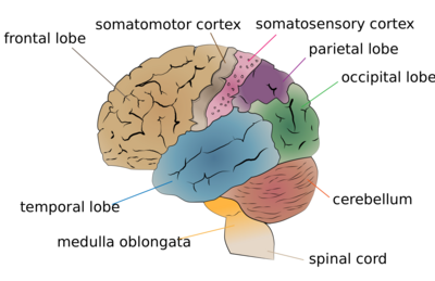
- Cerebrum: The largest part of the forebrain, responsible for higher brain functions such as thought, action, and sensory processing. The cerebral cortex, which is the outer layer of the cerebrum, is divided into four lobes:

Cerebrum lobes
- Frontal Lobe: Located underneath the forehead, it has three separate regions:
- Motor Cortex: Directs fine motor movements.
- Prefrontal Cortex: Involved with executive functioning, such as planning and decision-making.
- Broca’s Area: Crucial for speech production.
- Parietal Lobe: Located underneath the top rear part of the skull, it contains the somatosensory cortex, which processes sensory information such as pain and pressure.
- Occipital Lobe: Located at the back of the head, it processes visual information.
- Temporal Lobe: Located on the sides of the head, it processes auditory information and includes Wernicke’s area, important for language comprehension.
Localization of Function
Localization of function refers to the concept that different parts of the brain are responsible for specific behaviors or functions. Key examples include:
- Broca’s Area: Located in the frontal lobe, it is crucial for speech production. Damage to this area can result in Broca’s aphasia, characterized by difficulty in speaking but with relatively preserved comprehension.
- Wernicke’s Area: Located in the temporal lobe, it is important for language comprehension. Damage to this area can result in Wernicke’s aphasia, characterized by fluent but nonsensical speech and poor comprehension.
- Motor Cortex: Located in the frontal lobe, it is responsible for voluntary movements. Each region of the motor cortex controls different parts of the body.
- Somatosensory Cortex: Located in the parietal lobe, it processes sensory information from various parts of the body.
- Visual Cortex: Located in the occipital lobe, it processes visual information.
- Auditory Cortex: Located in the temporal lobe, it processes auditory information.
Lateralization of Function

The human brain is divided into two hemispheres–left and right. Scientists continue to explore how some cognitive functions tend to be dominated by one side or the other; that is, how they are lateralized. Right cerebral hemisphere Left cerebral hemisphere
Lateralization of function refers to the tendency for some neural functions or cognitive processes to be more dominant in one hemisphere than the other. Examples include:
- Left Hemisphere: Typically involved in language, analytical thinking, and logic. It controls the right side of the body.
- Right Hemisphere: Typically involved in spatial abilities, face recognition, visual imagery, and music. It controls the left side of the body.
While certain functions are more dominant in one hemisphere, both hemispheres constantly communicate through the corpus callosum, a bundle of neural fibers. About 95% of people show a specialization for language in the left hemisphere.
Split-Brain or Callosal Syndrome Overview
In the late 1950s, treatment for severe epilepsy sometimes involved severing the corpus callosum to stop the spread of seizures. Split-brain or callosal syndrome occurs when the corpus callosum, which connects the two hemispheres of the brain, is severed to some extent. This results in a type of disconnection syndrome characterized by a set of symptoms due to the disruption of communication between the brain’s hemispheres. The surgical procedure to achieve this (corpus callosotomy) involves cutting the corpus callosum and is typically a last-resort treatment for severe, refractory epilepsy. Initially, surgeons may perform partial callosotomies; if these are unsuccessful, a complete callosotomy might be done to reduce the severity and frequency of seizures and prevent injury. Before opting for surgery, epilepsy is generally treated with medication. Post-surgery, neuropsychological assessments are commonly conducted.
When the right and left hemispheres are separated, each hemisphere operates independently, with its own perceptions, concepts, and impulses. This can lead to unique and sometimes conflicting behaviors. For example, there was a case where a split-brain patient experienced one hand pulling up his pants while the other pulled them down. In another instance, the patient’s left hand acted aggressively towards his wife, while the right hand intervened to stop the left hand’s actions (a phenomenon known as alien hand syndrome). However, such conflicts are infrequent, and typically, one hemisphere’s actions override the other.
In split-brain patients, presenting an image to only the left visual field means they cannot verbally identify it. This is due to the contralateral nature of sensory processing, where the right hemisphere processes information from the left visual field. Without communication between hemispheres, the patient cannot articulate what the right hemisphere perceives. A similar effect occurs when a split-brain patient touches an object with only the left hand without visual cues from the right visual field; the patient cannot name the object. If the speech-control center is in the right hemisphere, presenting the object or image to the right visual field or hand will produce the same naming difficulty.
Brain-Imaging Techniques

An animated gif of MRI images of a human head.
Brain-imaging techniques are used to visualize the structure and function of the brain, aiding in the understanding of its complex workings. Key techniques include:
- Magnetic Resonance Imaging (MRI): Provides detailed images of brain anatomy using magnetic fields and radio waves. Functional MRI (fMRI) measures brain activity by detecting changes associated with blood flow.
- Computed Tomography (CT) Scan: Uses X-rays to create detailed images of the brain. It is useful for detecting brain injuries and tumors.
- Positron Emission Tomography (PET) Scan: Uses radioactive tracers to visualize brain activity by measuring changes in blood flow, oxygen use, and glucose metabolism.
- Electroencephalography (EEG): Measures electrical activity in the brain through electrodes placed on the scalp. It is useful for diagnosing conditions like epilepsy and for studying sleep and other states of consciousness.
- Magnetoencephalography (MEG): Measures the magnetic fields produced by neuronal activity in the brain. It provides detailed information about brain activity and is used in research and clinical diagnostics.
- Functional magnetic resonance imaging (fMRI) uses the magnetic properties of the blood to achieve the same goals without radiation.
- Other Brain-Recording TechniquesThe technique of event-related potential (ERP) measures an area of the brain’s response to a specific event.A newer technique, transcranial magnetic stimulation (TMS) allows investigators to measure activity of specific brain circuits when an area of the brain is excited or inhibited.
Summary
Understanding the structure and function of the brain involves exploring how different regions and hemispheres specialize in certain tasks (localization and lateralization) and utilizing various brain-imaging techniques to observe and measure brain activity. This comprehensive approach allows for a deeper understanding of how the brain controls behavior and processes information
Localization of Function and Franz Gall
The idea of localization of function, the concept that different parts of the brain are responsible for specific functions or behaviors, traces back to the work of the Austrian anatomist Franz Joseph Gall (1758-1828). Gall believed in faculty psychology, the theory that different mental abilities were carried out in different parts of the brain.
Gall’s student, Johan Spurzheim, developed the study of phrenology, the incorrect idea that differences in psychological abilities could be seen in the relative size of different brain areas.
Franz Joseph Gall and Phrenology

Franz Joseph Gall examining the head of a pretty young girl
- Background and Early Work:
- Franz Gall was a pioneer in the study of the brain and its functions. He believed that the brain was the organ of the mind and that different mental functions were localized in different areas of the brain.
- Gall’s interest in localization of function began with his observations of people around him. He noticed that individuals with pronounced abilities in certain areas (such as language or memory) often had specific parts of their skulls that seemed more developed.
- Phrenology:
- Gall developed a pseudoscientific theory called phrenology. According to this theory, the brain is composed of multiple, distinct “organs,” each responsible for different personality traits and intellectual capabilities.
- Gall proposed that the size and shape of these brain organs would affect the shape of the overlying skull. Thus, by examining the bumps and indentations on a person’s skull, one could infer their personality traits and mental abilities.
- In 1800, Johann Spurzheim attended one of Gall’s public lectures and was hired as an assistant to help with public medical demonstrations. In 1804, he became Gall’s full-time research partner.They worked together for years to develop theories about brain localization and function. In 1813, Spurzheim separated from Gall in order to make a name for himself in Britain. Gall would later accuse Spurzheim of plagiarism and perverting his work.It was Spurzheim who would give the name phrenology to Gall’s theories.
- Phrenological Map:
- Gall and his followers created detailed maps of the skull, marking various regions purportedly linked to different functions, such as combativeness, benevolence, and constructiveness.
- Despite being widely popular in the 19th century, phrenology was later discredited as a science because it lacked empirical support and rigorous scientific methodology.
- Impact on Neuroscience:
- While phrenology itself was flawed, Gall’s underlying idea that specific mental functions are localized in specific areas of the brain was influential. His work laid the groundwork for future neuroscientists to explore the relationship between brain structure and function.
- Gall’s emphasis on the brain as the seat of all mental activity contributed to the shift away from viewing the heart or other organs as the center of thought and emotion.
Advances Post-Gall
- Paul Broca and Broca’s Area:
- In the 19th century, French physician Paul Broca built on Gall’s ideas by providing clinical evidence for localization of function. He studied patients with speech impairments and discovered that damage to a specific area of the frontal lobe, now known as Broca’s area, resulted in difficulties in speech production.
- Broca’s work was crucial in establishing the concept of localization of function in the brain and demonstrating that specific brain regions have specific roles.
- Carl Wernicke and Wernicke’s Area:
- Following Broca’s findings, German neurologist Carl Wernicke identified another region of the brain associated with language comprehension, now known as Wernicke’s area. Damage to this area resulted in fluent but nonsensical speech and poor comprehension, a condition known as Wernicke’s aphasia.
- Wernicke’s work further reinforced the idea that different cognitive functions are localized in different parts of the brain.
- Modern Neuroscience:
- Today, the concept of localization of function is supported by a wealth of scientific evidence from various fields, including neuropsychology, neuroimaging, and cognitive neuroscience.
- Techniques such as fMRI and PET scans allow researchers to observe brain activity in real time, providing insights into how specific areas of the brain contribute to different mental processes and behaviors.
Summary
Franz Joseph Gall’s early work on localization of function, despite being embedded in the flawed science of phrenology, was pivotal in advancing the idea that different regions of the brain are responsible for different functions. This idea was later validated and refined by subsequent research, leading to our current understanding of brain localization and its crucial role in cognitive neuroscience.
Training the Brain
Research with young animals suggests that experience can significantly alter the brain’s structure. For instance, studies with rats have shown that those raised in enriched environments—featuring a variety of toys, activities, and social interactions—demonstrate better brain development compared to rats raised in standard, less stimulating cages. These enriched environments appear to enhance neuronal growth, increase synaptic connections, and improve overall cognitive function in these animals.
When it comes to human adults, the results of “brain training games” on cognitive functioning have been mixed. While some studies suggest that these games can lead to improvements in specific cognitive skills, such as memory or attention, other research indicates that these benefits do not always generalize to broader cognitive abilities or everyday tasks. Some researchers argue that while brain training games can improve performance on the tasks practiced within the games, they do not necessarily translate to improved cognitive functioning in real-world scenarios. As a result, the effectiveness of brain training as a tool for enhancing overall cognitive health remains a topic of ongoing research and debate.
Brain Plasticity and Ablation Studies
Further evidence of the brain’s adaptability comes from Lashley’s studies of ablation, where parts of the brain were surgically removed in rats. His research showed that the rats’ abilities to navigate mazes were related to the total amount of cortex removed, rather than the specific area. This finding suggests that the brain functions as a whole and that multiple regions can contribute to complex behaviors, such as maze running.
Additionally, the concept of brain plasticity highlights the brain’s remarkable ability to reorganize itself, especially in response to injury. This phenomenon is particularly pronounced in younger individuals, where undamaged brain regions can take over the functions of damaged areas. Plasticity underscores the brain’s capacity for adaptation and recovery, illustrating that neural pathways can be reshaped by experience and learning.
Key Takeaways
- Brain Plasticity: The brain’s ability to reorganize itself by forming new neural connections, especially in response to injury or experience.
- Ablation: The surgical removal of brain tissue used in research to study the effects on behavior and brain function.
- Enriched Environments: Settings that provide enhanced sensory, cognitive, and social stimulation, leading to better brain development in animals.
- Brain Training Games: Games designed to improve cognitive functions such as memory, attention, and problem-solving skills.
- Split-Brain Syndrome: A condition resulting from the severing of the corpus callosum, leading to the independent functioning of the brain’s hemispheres.
- Corpus Callosotomy: A surgical procedure that involves cutting the corpus callosum to treat severe epilepsy.
- Neuropsychological Assessments: Evaluations used to measure cognitive functioning and brain behavior relationships, often conducted after brain surgeries.
- Contralateral Processing: The brain’s method of processing sensory information on the opposite side of the body from where the information was received.
- Alien Hand Syndrome: A phenomenon where one hand acts independently of conscious control, sometimes observed in split-brain patients.
- Cerebral Cortex: The outer layer of the brain, involved in complex functions such as thought, perception, and voluntary movement.
- Thalamus: A brain structure that relays sensory and motor signals to the cerebral cortex.
- Hypothalamus: A brain region that regulates various physiological functions by releasing hormones.
- Hippocampus: A brain region involved in the formation of long-term memories.
- Amygdala: A brain structure involved in emotional learning and memory.
- Frontal Lobe: A region of the cerebral cortex involved in executive functions, motor control, and decision-making.
- Parietal Lobe: A region of the cerebral cortex that processes sensory information such as pain and pressure.
- Occipital Lobe: The part of the cerebral cortex that processes visual information.
- Temporal Lobe: The region of the cerebral cortex involved in processing auditory information.
- Localization of Function: The concept that specific areas of the brain are responsible for specific functions.
- Lateralization of Function: The tendency for some neural functions to be more dominant in one hemisphere than the other.
Candela Citations
Public domain content
- Image Franz Joseph Gall examining the head of a pretty young girl. Authored by: By Possibly Edward Hull . Located at: https://en.wikipedia.org/wiki/Franz_Joseph_Gall#/media/File:Franz_Joseph_Gall_examining_the_head_of_a_pretty_young_girl,_Wellcome_V0011119.jpg. License: CC BY: Attribution. License Terms: By Possibly Edward Hull - https://wellcomeimages.org/indexplus/image/V0011119.htmlWellcome Collection gallery (2018-03-29): https://wellcomecollection.org/works/m2utphh3 CC-BY-4.0, CC BY 2.0, https://commons.wikimedia.org/w/index.php?curid=31233962
- Franz Gall. Provided by: Wikipedia Foundation, Inc.. Located at: https://en.wikipedia.org/wiki/Franz_Joseph_Gall. License: CC BY-SA: Attribution-ShareAlike. License Terms: Text is available under the Creative Commons Attribution-ShareAlike License 4.0; additional terms may apply. By using this site, you agree to the Terms of Use and Privacy Policy. Wikipediau00ae is a registered trademark of the Wikimedia Foundation, Inc., a non-profit organization.
- Cerebral hemispheres . Authored by: By Polygon data were generated by Database Center for Life Science (DBCLS). Provided by: Wikipedia.org. Located at: https://en.wikipedia.org/wiki/Lateralization_of_brain_function#/media/File:Cerebral_hemisphere_-_animation.gif. License: CC BY-SA: Attribution-ShareAlike. License Terms: By Polygon data were generated by Database Center for Life Science (DBCLS) - Polygon data are from BodyParts3D, CC BY-SA 2.1 jp, https://commons.wikimedia.org/w/index.php?curid=79950202
- Picture Cerebrum lobes. Authored by: By Vectorized image by Jkwchui, via Wikimedia Commons. Labeled for reuse. - https://upload.wikimedia.org/wikipedia/commons/thumb/d/d8/Cerebrum_lobes.svg/2000px-Cerebrum_lobes.svg.png, CC BY-SA 4.0, https://commons.wikimedia.org/w/index.php?curid=51205157. Located at: https://en.wikipedia.org/wiki/Perception. License: CC BY-SA: Attribution-ShareAlike
Lumen Learning authored content
- Use of ChatGPT to detail learning objectives. Authored by: Sonja Miller. Provided by: Hudson Valley Community College. Project: Creation of OER for Cognitive Psychology. License: CC BY-SA: Attribution-ShareAlike


