Learning Objectives
By the end of this section, you will be able to:
- Describe the structural organization of nematodes
- Understand the importance of Caenorhabditis elegans in research
- Compare the internal systems and appendage specializations of phylum Arthropoda
- Discuss the environmental importance of arthropods
- Discuss the reasons for arthropod success and abundance
The superphylum Ecdysozoa contains an incredibly large number of species. This is because it contains two of the most diverse animal groups: phylum Nematoda (the roundworms) and Phylum Arthropoda (the arthropods). The most prominant distinguising feature of Ecdysozoans is their tough external covering called the cuticle. The cuticle provides a tough, but flexible exoskeleton tht protects these animals from water loss, predators and other aspects of the external environment. All members of this superphylum periodically molt, or shed their cuticle as they grow. After molting, they secrete a new cuticle that will last until their next growth phase. The process of molting and replacing the cuticle is called ecdysis, which is how the superphylum derived its name.
Phylum Nematoda
The Nematoda, like most other animal phyla, are triploblastic and possess an embryonic mesoderm that is sandwiched between the ectoderm and endoderm. They are also bilaterally symmetrical, meaning that a longitudinal section will divide them into right and left sides that are symmetrical. Furthermore, the nematodes, or roundworms, possess a pseudocoelom and consist of both free-living and parasitic forms.
It has been said that were all the non-nematode matter of the biosphere removed, there would remain a shadow of the former world in the form of nematodes.[1] The arthropods, one of the most successful taxonomic groups on the planet, are coelomate organisms characterized by a hard exoskeleton and jointed appendages. Both the nematodes and arthropods belong to the superphylum Ecdysozoa that is believed to be a clade consisting of all evolutionary descendants from one common ancestor. The name derives from the word ecdysis, which refers to the shedding, or molting, of the exoskeleton. The phyla in this group have a hard cuticle that covers their bodies, which must be periodically shed and replaced for them to increase in size.
Phylum Nematoda includes more than 28,000 species with an estimated 16,000 being parasitic in nature. The name Nematoda is derived from the Greek word “Nemos,” which means “thread” and includes roundworms. Nematodes are present in all habitats with a large number of individuals of each species present in each. The free-living nematode, Caenorhabditis elegans has been extensively used as a model system in laboratories all over the world.
Morphology
In contrast with cnidarians, nematodes show a tubular morphology and circular cross-section. These animals are pseudocoelomates and show the presence of a complete digestive system with a distinct mouth and anus. This is in contrast with the cnidarians, where only one opening is present (an incomplete digestive system).
The cuticle of Nematodes is rich in collagen and a carbohydrate-protein polymer called chitin, and forms an external “skeleton” outside the epidermis. The cuticle also lines many of the organs internally, including the pharynx and rectum. The epidermis can be either a single layer of cells or a syncytium, which is a multinucleated cell formed from the fusion of uninucleated cells.
The overall morphology of these worms is cylindrical, as seen in Figure 1. The head is radially symmetrical. A mouth opening is present at the anterior end with three or six lips as well as teeth in some species in the form of cuticle extensions. Some nematodes may present other external modifications like rings, head shields, or warts. Rings, however, do not reflect true internal body segmentation. The mouth leads to a muscular pharynx and intestine, which leads to a rectum and anal opening at the posterior end. The muscles of nematodes differ from those of most animals: They have a longitudinal layer only, which accounts for the whip-like motion of their movement.

Figure 1. Scanning electron micrograph shows (a) the soybean cyst nematode (Heterodera glycines) and a nematode egg. (b) A schematic representation shows the anatomy of a typical nematode. (credit a: modification of work by USDA ARS; scale-bar data from Matt Russell)
Excretory System
In nematodes, specialized excretory systems are not well developed. Nitrogenous wastes may be lost by diffusion through the entire body or into the pseudocoelom (body cavity), where they are removed by specialized cells. Regulation of water and salt content of the body is achieved by renette glands, present under the pharynx in marine nematodes.
Nervous system
Most nematodes possess four longitudinal nerve cords that run along the length of the body in dorsal, ventral, and lateral positions. The ventral nerve cord is better developed than the dorsal or lateral cords. All nerve cords fuse at the anterior end, around the pharynx, to form head ganglia or the “brain” of the worm (which take the form of a ring around the pharynx) as well as at the posterior end to form the tail ganglia. In C. elegans, the nervous system accounts for nearly one-third of the total number of cells in the animal.
Reproduction
Nematodes employ a variety of reproductive strategies that range from monoecious to dioecious to parthenogenic, depending upon the species under consideration. C. elegans is a monoecious species and shows development of ova contained in a uterus as well as sperm contained in the spermatheca. The uterus has an external opening known as the vulva. The female genital pore is near the middle of the body, whereas the male’s is at the tip. Specialized structures at the tail of the male keep him in place while he deposits sperm with copulatory spicules. Fertilization is internal, and embryonic development starts very soon after fertilization. The embryo is released from the vulva during the gastrulation stage. The embryonic development stage lasts for 14 hours; development then continues through four successive larval stages with ecdysis between each stage—L1, L2, L3, and L4—ultimately leading to the development of a young male or female adult worm. Adverse environmental conditions like overcrowding and lack of food can result in the formation of an intermediate larval stage known as the dauer larva.
Everyday Connection
C. elegans: The Model System for Linking Developmental Studies with GeneticsIf biologists wanted to research how nicotine dependence develops in the body, how lipids are regulated, or observe the attractant or repellant properties of certain odors, they would clearly need to design three very different experiments. However, they might only need one object of study: C. elegans. The nematode Caenorhabditis elegans was brought into the focus of mainstream biological research by Dr. Sydney Brenner. Since 1963, Dr. Brenner and scientists worldwide have used this animal as a model system to study various physiological and developmental mechanisms.
C. elegans is a free-living organism found in soil. It is easy to culture this organism on agar plates (10,000 worms/plate), it feeds on Escherichia coli (another long-term resident of biological laboratories worldwide), and therefore, it can be readily grown and maintained in a laboratory. The biggest asset of this nematode is its transparency, which helps researchers to observe and monitor changes within the animal with ease. It is also a simple organism with fewer than 1,000 cells and a genome of 20,000 genes. It shows chromosomal organization of DNA into five pairs of autosomes plus a pair of sex chromosomes, making it an ideal candidate to study genetics. Since every cell can be visualized and identified, this organism is useful for studying cellular phenomena like cell-cell interactions, cell-fate determinations, cell division, apoptosis, and intracellular transport.
Another tremendous asset is the short life cycle of this worm (Figure 2). It takes only 3 days to achieve the “egg to adult to daughter egg;” therefore, tracking genetic changes is easier in this animal. The total life span of C. elegans is 2 to 3 weeks; hence, age-related phenomena are easy to observe. Another feature that makes C. elegans an excellent experimental model system is that the position and number of the 959 cells present in adult hermaphrodites of this organism is constant. This feature is extremely significant when studying cell differentiation, cell-cell communication, and apoptosis. Lastly, C. elegans is also amenable to genetic manipulations using molecular methods, rounding off its usefulness as a model system.
Biologists worldwide have created information banks and groups dedicated to research using C. elegans. Their findings have led, for example, to better understandings of cell communication during development, neuronal signaling and insight into lipid regulation (which is important in addressing health issues like the development of obesity and diabetes). In recent years, studies have enlightened the medical community with a better understanding of polycystic kidney disease. This simple organism has led biologists to complex and significant findings, growing the field of science in ways that touch the everyday world.

Figure 2. (a) This light micrograph shows Caenorhabditis elegans. Its transparent adult stage consists of exactly 959 cells. (b) The life cycle of C. elegans has four juvenile stages (L1 through L4) and an adult stage. Under ideal conditions, the nematode spends a set amount of time at each juvenile stage, but under stressful conditions, it may enter a dauer state that does not age. The worm is hermaphroditic in the adult state, and mating of two worms produces a fertilized egg. (credit a: modification of work by “snickclunk”/Flickr: credit b: modification of work by NIDDK, NIH; scale-bar data from Matt Russell)
A number of common parasitic nematodes serve as prime examples of parasitism. These animals exhibit complex lifecycles that involve multiple hosts, and they can have significant medical and veterinary impacts. Humans may become infected by Dracunculus medinensis, known as guinea worms, when they drink unfiltered water containing copepods (Figure 3). Hookworms, such as Ancyclostoma and Necator, infest the intestines and feed on the blood of mammals, especially in dogs, cats, and humans. Trichina worms (Trichinella) are the causal organism of trichinosis in humans, often resulting from the consumption of undercooked pork; Trichinella can infect other mammalian hosts as well. Ascaris, a large intestinal roundworm, steals nutrition from its human host and may create physical blockage of the intestines. The filarial worms, such as Dirofilaria and Wuchereria, are commonly vectored by mosquitoes, which pass the infective agents among mammals through their blood-sucking activity. Dirofilaria immitis, a blood-infective parasite, is the notorious dog heartworm species. Wuchereria bancrofti infects the lymph nodes of humans, resulting in the non-lethal but deforming condition called elephantiasis, in which parts of the body become swelled to gigantic proportions due to obstruction of lymphatic drainage and inflammation of lymphatic tissues.

Figure 3. The guinea worm Dracunculus medinensis infects about 3.5 million people annually, mostly in Africa. (a) Here, the worm is wrapped around a stick so it can be extracted. (b) Infection occurs when people consume water contaminated by infected copepods, but this can easily be prevented by simple filtration systems. (credit: modification of work by CDC)
Phylum Arthropoda
The name “arthropoda” means “jointed legs” (in the Greek, “arthros” means “joint” and “podos” means “leg”); it aptly describes the enormous number of invertebrates included in this phylum. Arthropoda dominate the animal kingdom with an estimated 85 percent of known species included in this phylum and many arthropods yet undocumented. The principal characteristics of all the animals in this phylum are functional segmentation of the body and presence of jointed appendages. Arthropods also show the presence of an exoskeleton made principally of chitin, which is a waterproof, tough polysaccharide. Phylum Arthropoda is the largest phylum in the animal world, and insects form the single largest class within this phylum. Arthropods are eucoelomate, protostomic organisms.

Figure 4. Trilobites, like the one in this fossil, are an extinct group of arthropods. (credit: Kevin Walsh)
Phylum Arthropoda includes animals that have been successful in colonizing terrestrial, aquatic, and aerial habitats. This phylum is further classified into five subphyla: Trilobitomorpha (trilobites, all extinct), Hexapoda (insects and relatives), Myriapoda (millipedes, centipedes, and relatives), Crustaceans (crabs, lobsters, crayfish, isopods, barnacles, and some zooplankton), and Chelicerata (horseshoe crabs, arachnids, scorpions, and daddy longlegs). Trilobites are an extinct group of arthropods found chiefly in the pre-Cambrian Era that are probably most closely related to the Chelicerata. These are identified based on fossil records (Figure 4).
Morphology
A unique feature of animals in the arthropod phylum is the presence of a segmented body and fusion of sets of segments that give rise to functional body regions called tagma. Tagma may be in the form of a head, thorax, and abdomen, or a cephalothorax and abdomen, or a head and trunk. A central cavity, called the hemocoel (or blood cavity), is present, and the open circulatory system is regulated by a tubular or single-chambered heart. Respiratory systems vary depending on the group of arthropod: insects and myriapods use a series of tubes (tracheae) that branch through the body, open to the outside through openings called spiracles, and perform gas exchange directly between the cells and air in the tracheae, whereas aquatic crustaceans utilize gills, terrestrial chelicerates employ book lungs, and aquatic chelicerates use book gills (Figure 5). The book lungs of arachnids (scorpions, spiders, ticks and mites) contain a vertical stack of hemocoel wall tissue that somewhat resembles the pages of a book. Between each of the “pages” of tissue is an air space. This allows both sides of the tissue to be in contact with the air at all times, greatly increasing the efficiency of gas exchange. The gills of crustaceans are filamentous structures that exchange gases with the surrounding water. Groups of arthropods also differ in the organs used for excretion, with crustaceans possessing green glands and insects using Malpighian tubules, which work in conjunction with the hindgut to reabsorb water while ridding the body of nitrogenous waste. The cuticle is the covering of an arthropod. It is made up of two layers: the epicuticle, which is a thin, waxy water-resistant outer layer containing no chitin, and the layer beneath it, the chitinous procuticle. Chitin is a tough, flexible polysaccharide. In order to grow, the arthropod must shed the exoskeleton during a process called ecdysis (“to strip off”); this is a cumbersome method of growth, and during this time, the animal is vulnerable to predation. The characteristic morphology of representative animals from each subphylum is described below.

Figure 5. The book lungs of (a) arachnids are made up of alternating air pockets and hemocoel tissue shaped like a stack of books. The book gills of (b) crustaceans are similar to book lungs but are external so that gas exchange can occur with the surrounding water. (credit a: modification of work by Ryan Wilson based on original work by John Henry Comstock; credit b: modification of work by Angel Schatz)
Subphylum Hexapoda
The name Hexapoda denotes the presence of six legs (three pairs) in these animals as differentiated from the number of pairs present in other arthropods. Hexapods are characterized by the presence of a head, thorax, and abdomen, constituting three tagma. The thorax bears the wings as well as six legs in three pairs. Many of the common insects we encounter on a daily basis—including ants, cockroaches, butterflies, and flies—are examples of Hexapoda.
Amongst the hexapods, the insects (Figure 6) are the largest class in terms of species diversity as well as biomass in terrestrial habitats. Typically, the head bears one pair of sensory antennae, mandibles as mouthparts, a pair of compound eyes, and some ocelli (simple eyes) along with numerous sensory hairs. The thorax bears three pairs of legs (one pair per segment) and two pairs of wings, with one pair each on the second and third thoracic segments. The abdomen usually has eleven segments and bears reproductive apertures. Hexapoda includes insects that are winged (like fruit flies) and wingless (like fleas).
Art Connection

Figure 6. In this basic anatomy of a hexapod insect, note that insects have a developed digestive system (yellow), a respiratory system (blue), a circulatory system (red), and a nervous system (red).
Which of the following statements about insects is false?
- Insects have both dorsal and ventral blood vessels.
- Insects have spiracles, openings that allow air to enter.
- The trachea is part of the digestive system.
- Insects have a developed digestive system with a mouth, crop, and intestine.
Figure C is false.
Subphylum Myriapoda
Subphylum Myriapoda includes arthropods with numerous legs. Although the name is hyperbolic in suggesting that myriad legs are present in these invertebrates, the number of legs may vary from 10 to 750. This subphylum includes 13,000 species; the most commonly found examples are millipedes and centipedes. All myriapods are terrestrial animals and prefer a humid environment.
Myriapods are typically found in moist soils, decaying biological material, and leaf litter. Subphylum Myriapoda is divided into four classes: Chilopoda, Symphyla, Diplopoda, and Pauropoda. Centipedes like Scutigera coleoptrata (Figure 7a) are classified as chilopods. These animals bear one pair of legs per segment, mandibles as mouthparts, and are somewhat dorsoventrally flattened. The legs in the first segment are modified to form forcipules (poison claws) that deliver poison to prey like spiders and cockroaches, as these animals are all predatory. Millipedes bear two pairs of legs per diplosegment, a feature that results from embryonic fusion of adjacent pairs of body segments, are usually rounder in cross-section, and are herbivores or detritivores. Millipedes have visibly more numbers of legs as compared to centipedes, although they do not bear a thousand legs (Figure 7b).
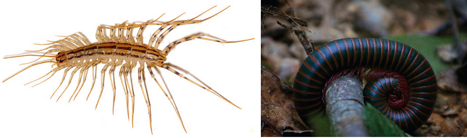
Figure 7. (a) The Scutigera coleoptrata centipede has up to 15 pairs of legs. (b) This North American millipede (Narceus americanus) bears many legs, although not a thousand, as its name might suggest. (credit a: modification of work by Bruce Marlin; credit b: modification of work by Cory Zanker)
Subphylum Crustacea
Crustaceans are the most dominant aquatic arthropods, since the total number of marine crustacean species stands at 67,000, but there are also freshwater and terrestrial crustacean species. Krill, shrimp, lobsters, crabs, and crayfish are examples of crustaceans (Figure 8). Terrestrial species like the wood lice (Armadillidium spp.) (also called pill bugs, rolly pollies, potato bugs, or isopods) are also crustaceans, although the number of non-aquatic species in this subphylum is relatively low.

Figure 8. The (a) crab and (b) shrimp krill are both crustaceans. (credit a: modification of work by William Warby; credit b: modification of work by Jon Sullivan)
Crustaceans possess two pairs of antennae, mandibles as mouthparts, and biramous (“two branched”) appendages, which means that their legs are formed in two parts, as distinct from the uniramous (“one branched”) myriapods and hexapods (Figure 9).

Figure 9. Arthropods may have (a) biramous (two-branched) appendages or (b) uniramous (one-branched) appendages. (credit b: modification of work by Nicholas W. Beeson)
Unlike that of the Hexapoda, the head and thorax of most crustaceans is fused to form a cephalothorax (Figure 10), which is covered by a plate called the carapace, thus producing a body structure of two tagma. Crustaceans have a chitinous exoskeleton that is shed by molting whenever the animal increases in size. The exoskeletons of many species are also infused with calcium carbonate, which makes them even stronger than in other arthropods. Crustaceans have an open circulatory system where blood is pumped into the hemocoel by the dorsally located heart. Hemocyanin and hemoglobin are the respiratory pigments present in these animals.

Figure 10. The crayfish is an example of a crustacean. It has a carapace around the cephalothorax and the heart in the dorsal thorax area. (credit: Jane Whitney)
Most crustaceans are dioecious, which means that the sexes are separate. Some species like barnacles may be hermaphrodites. Serial hermaphroditism, where the gonad can switch from producing sperm to ova, may also be seen in some species. Fertilized eggs may be held within the female of the species or may be released in the water. Terrestrial crustaceans seek out damp spaces in their habitats to lay eggs.
Larval stages—nauplius and zoea—are seen in the early development of crustaceans. A cypris larva is also seen in the early development of barnacles (Figure 11).

Figure 11. All crustaceans go through different larval stages. Shown are (a) the nauplius larval stage of a tadpole shrimp, (b) the cypris larval stage of a barnacle, and (c) the zoea larval stage of a green crab. (credit a: modification of work by USGS; credit b: modification of work by Mª. C. Mingorance Rodríguez; credit c: modification of work by B. Kimmel based on original work by Ernst Haeckel)
Crustaceans possess a tripartite brain and two compound eyes. Most crustaceans are carnivorous, but herbivorous and detritivorous species are also known. Crustaceans may also be cannibalistic when extremely high populations of these organisms are present.
Subphylum Chelicerata
This subphylum includes animals such as spiders, scorpions, horseshoe crabs, and sea spiders. This subphylum is predominantly terrestrial, although some marine species also exist. An estimated 77,000 species are included in subphylum Chelicerata. Chelicerates are found in almost all habitats.

Figure 12. The chelicerae (first set of appendages) are well developed in the scorpion. (credit: Kevin Walsh)
The body of chelicerates may be divided into two parts: prosoma and opisthosoma, which are basically the equivalents of cephalothorax (usually smaller) and abdomen (usually larger). A “head” tagmum is not usually discernible. The phylum derives its name from the first pair of appendages: the chelicerae (Figure 12), which are specialized, claw-like or fang-like mouthparts. These animals do not possess antennae. The second pair of appendages is known as pedipalps. In some species, like sea spiders, an additional pair of appendages, called ovigers, is present between the chelicerae and pedipalps.
Chelicerae are mostly used for feeding, but in spiders, these are often modified into fangs that inject venom into their prey before feeding (Figure 13). Members of this subphylum have an open circulatory system with a heart that pumps blood into the hemocoel. Aquatic species have gills, whereas terrestrial species have either trachea or book lungs for gaseous exchange.

Figure 13. The trapdoor spider, like all spiders, is a member of the subphylum Chelicerata. (credit: Marshal Hedin)
Most chelicerates ingest food using a preoral cavity formed by the chelicerae and pedipalps. Some chelicerates may secrete digestive enzymes to pre-digest food before ingesting it. Parasitic chelicerates like ticks and mites have evolved blood-sucking apparatuses.
The nervous system in chelicerates consists of a brain and two ventral nerve cords. These animals use external fertilization as well as internal fertilization strategies for reproduction, depending upon the species and its habitat. Parental care for the young ranges from absolutely none to relatively prolonged care.
Link to Learning
Visit this site to click through a lesson on arthropods, including interactive habitat maps, and more.
Section Summary
Nematodes are pseudocoelomate animals akin to flatworms, yet display more advanced neuronal development, a complete digestive system, and a body cavity. This phylum includes free-living as well as parasitic organisms like Caenorhabditis elegans and Ascaris spp., respectively. They include dioeceous as well as hermaphroditic species. Nematodes also possess an excretory system that is not quite well developed. Embryonic development is external and proceeds via three larval stages. A peculiar feature of nematodes is the secretion of a collagenous/chitinous cuticle outside the body.
Arthropods represent the most successful phylum of animal on Earth, in terms of the number of species as well as the number of individuals. These animals are characterized by a segmented body as well as the presence of jointed appendages. In the basic body plan, a pair of appendages is present per body segment. Within the phylum, traditional classification is based on mouthparts, number of appendages, and modifications of appendages present. Arthropods bear a chitinous exoskeleton. Gills, trachea, and book lungs facilitate respiration. Sexual dimorphism is seen in this phylum, and embryonic development includes multiple larval stages.
Candela Citations
- Biology. Authored by: OpenStax. Provided by: OpenStax College. Located at: http://cnx.org/contents/185cbf87-c72e-48f5-b51e-f14f21b5eabd@9.44:1/Biology. License: CC BY: Attribution
- Stoll, N. R., “This wormy world. 1947,” Journal of Parasitology 85(3) (1999): 392–96. ↵