Describe the circulatory, immune, respiratory, and digestive systems
This set of body systems has been grouped together as the “cell maintenance systems.” Remember, this isn’t a hard-and-fast categorization: these systems are grouped together to help you organize your learning. These body systems all function to maintain the cells in your body: the circulatory, respiratory, and digestive systems provide the nutrients and energy your cells need to live. The immune system protects these cells from pathogens.
Learning Objectives
- Identify the structure and function of the circulatory system
- Identify the structure and function of the respiratory system
- Identify the structure and function of the immune system
- Identify the structure and function of the digestive system
Circulatory System
The circulatory system is extremely important in sustaining life. It’s proper functioning is responsible for the delivery of oxygen and nutrients to all cells, as well as the removal of carbon dioxide, waste products, maintenance of optimum pH, and the mobility of the elements, proteins and cells, of the immune system. In developed countries, the two leading causes of death, myocardial infarction and stroke are each direct results of an arterial system that has been slowly and progressively compromised by years of deterioration. The circulatory system includes the heart, blood vessels, blood, lymph and lymph vessels. But we often think of it as the vascular network that connects to the primary cardiac organ, the heart. More accurately the vascular network directly connected to the heart is just blood and blood vessels, but do not be surprised to see the terms circulatory and cardiovascular used interchangeably. Here we are focusing on the heart and vascular network that transports blood.
The Heart

Figure 1. Blood vessels of the coronary system, including the coronary arteries and veins, keep the heart musculature oxygenated.
The heart is the life-giving, ever-beating muscle in your chest. From inside the womb until death, the thump goes on. The heart for the average human will contract about 3 billion times; never resting, never stopping to take a break except for a fraction of a second between beats. At 80 years of age, a person’s heart will continue to beat an average of 100,000 times a day. Many believe that the heart is the first organ to become functional. Within weeks of conception the heart starts its mission of supplying the body with nutrients even though the embryo is no bigger than a capital letter on this page. The primary function of the heart is to pump blood through the arteries, capillaries, and veins. There are an estimated 60,000 miles of vessels throughout an adult body. Blood transports oxygen, nutrients, disease causing viruses, bacteria, hormones and has other important functions as well. The heart is the pump that keeps blood circulating properly. Americans today have many options to take care of their heart and circulatory system. Expanding medical technology has made it much easier to do so.
The heart is a hollow, muscular organ about the size of a fist. It is responsible for pumping blood through the blood vessels by repeated, rhythmic contractions. The heart is composed of cardiac muscle, an involuntary muscle tissue that is found only within this organ. The term “cardiac” (as in cardiology) means “related to the heart” and comes from the Greek word kardia, for “heart.” It has a four-chambered, double pump and is located in the thoracic cavity between the lungs. The cardiac muscle is self-exciting, meaning it has its own conduction system. This is in contrast with skeletal muscle, which requires either conscious or reflex nervous stimuli. The heart’s rhythmic contractions occur spontaneously, although the frequency or heart rate can be changed by nervous or hormonal influence such as exercise or the perception of danger.
The Cardiovascular System
Arteries
Arteries are muscular blood vessels that carry blood away from the heart, oxygenated and deoxygenated blood. The pulmonary arteries will carry deoxygenated blood to the lungs and the systemic arteries will carry oxygenated blood to the rest of the body. Arteries have a thick wall that consists of three layers. The inside layer is called the endothelium, the middle layer is mostly smooth muscle and the outside layer is connective tissue. The artery walls are thick so that when blood enters under pressure the walls can expand.
Arterioles
An arteriole is a small artery that extends and leads to capillaries. Arterioles have thick smooth muscular walls. These smooth muscles are able to contract (causing vessel constriction) and relax (causing vessel dilation). This contracting and relaxing affects blood pressure; the higher number of vessels dilated, the lower blood pressure will be. Arterioles are just visible to the naked eye.
Capillaries

Figure 2. Capillaries
Capillaries are the smallest of a body’s vessels; they connect arteries and veins, and most closely interact with tissues. They are very prevalent in the body; total surface area is about 6,300 square meters. Because of this, no cell is very far from a capillary, no more than 50 micrometers away. The walls of capillaries are composed of a single layer of cells, the endothelium, which is the inner lining of all the vessels. This layer is so thin that molecules such as oxygen, water and lipids can pass through them by diffusion and enter the tissues. Waste products such as carbon dioxide and urea can diffuse back into the blood to be carried away for removal from the body.
The “capillary bed” is the network of capillaries present throughout the body. These beds are able to be “opened” and “closed” at any given time, according to need. This process is called autoregulation and capillary beds usually carry no more than 25 percent of the amount of blood it could hold at any time. The more metabolically active the cells, the more capillaries it will require to supply nutrients.
Veins
Veins carry blood to the heart. The pulmonary veins will carry oxygenated blood to the heart awhile the systemic veins will carry deoxygenated to the heart. Most of the blood volume is found in the venous system; about 70% at any given time. The veins outer walls have the same three layers as the arteries, differing only because there is a lack of smooth muscle in the inner layer and less connective tissue on the outer layer. Veins have low blood pressure compared to arteries and need the help of skeletal muscles to bring blood back to the heart. Most veins have one-way valves called venous valves to prevent backflow caused by gravity. They also have a thick collagen outer layer, which helps maintain blood pressure and stop blood pooling. If a person is standing still for long periods or is bedridden, blood can accumulate in veins and can cause varicose veins. The hollow internal cavity in which the blood flows is called the lumen. A muscular layer allows veins to contract, which puts more blood into circulation. Veins are used medically as points of access to the blood stream, permitting the withdrawal of blood specimens (venipuncture) for testing purposes, and enabling the infusion of fluid, electrolytes, nutrition, and medications (intravenous delivery).
Venules
A venule is a small vein that allows deoxygenated blood to return from the capillary beds to the larger blood veins, except in the pulmonary circuit were the blood is oxygenated. Venules have three layers; they have the same makeup as arteries with less smooth muscle, making them thinner.
Video Review
Watch this video for a quick survey of the human circulatory system:
Respiratory System
The Respiratory System is vital to every human being. Without it, we would cease to live outside of the womb. Let us begin by taking a look at the structure of the respiratory system and how vital it is to life. During inhalation or exhalation air is pulled towards or away from the lungs, by several cavities, tubes, and openings.
The organs of the respiratory system make sure that oxygen enters our bodies and carbon dioxide leaves our bodies.
The respiratory tract is the path of air from the nose to the lungs. It is divided into two sections: Upper Respiratory Tract and the Lower Respiratory Tract. Included in the upper respiratory tract are the Nostrils, Nasal Cavities, Pharynx, Epiglottis, and the Larynx. The lower respiratory tract consists of the Trachea, Bronchi, Bronchioles, and the Lungs.
As air moves along the respiratory tract it is warmed, moistened and filtered.

Figure 3. Click for a larger image. The major respiratory structures span the nasal cavity to the diaphragm.
Functions
There are four processes of respiration. They are:
- Breather or ventilation
- External Respiration, which is the exchange of gases (oxygen and carbon dioxide) between inhaled air and the blood.
- Internal Respiration, which is the exchange of gases between the blood and tissue fluids.
- Cellular Respiration
In addition to these main processes, the respiratory system serves for:
- Regulation of Blood pH, which occurs in coordination with the kidneys,
- Defense against microbes
- Control of body temperature due to loss of evaporate during expiration
Respiratory System: Upper and Lower Respiratory Tracts
For the sake of convenience, we will divide the respiratory system in to the upper and lower respiratory tracts:
Upper Respiratory Tract
The upper respiratory tract, can refer to the parts of the respiratory system lying above the sternal angle (outside of the thorax), above the vocal folds, or above the cricoid cartilage. The tract consists of the nasal cavity and paranasal sinuses, the pharynx (nasopharynx, oropharynx and laryngopharynx) and sometimes includes the larynx. Its primary function is to receive the air from the external environment and filter, warm, and humidify it before it reaches the delicate lungs where gas exchange will occur.
Air enters through the nostrils of the nose and is partially filtered by the nose hairs, then flows into the nasal cavity. The nasal cavity is lined with epithelial tissue, containing blood vessels, which help warm the air; and secrete mucous, which further filters the air. The endothelial lining of the nasal cavity also contains tiny hairlike projections, called cilia. The cilia serve to transport dust and other foreign particles, trapped in mucous, to the back of the nasal cavity and to the pharynx. There the mucus is either coughed out, or swallowed and digested by powerful stomach acids. After passing through the nasal cavity, the air flows down the pharynx to the larynx.
Lower Respiratory Tract
The lower respiratory tract or lower airway is derived from the developing foregut and consists of the trachea, bronchi (primary, secondary and tertiary), bronchioles (including terminal and respiratory), and lungs (including alveoli). It also sometimes includes the larynx, which we have done here. This is where gas exchange actually takes place.
Larynx
The larynx (plural larynges), colloquially known as the voice box, is an organ in our neck involved in protection of the trachea and sound production. The larynx houses the vocal cords, and is situated just below where the tract of the pharynx splits into the trachea and the esophagus. The larynx contains two important structures: the epiglottis and the vocal cords.
The epiglottis is a flap of cartilage located at the opening to the larynx. During swallowing, the larynx (at the epiglottis and at the glottis) closes to prevent swallowed material from entering the lungs; the larynx is also pulled upwards to assist this process. Stimulation of the larynx by ingested matter produces a strong cough reflex to protect the lungs. Note: choking occurs when the epiglottis fails to cover the trachea, and food becomes lodged in our windpipe.
The vocal cords consist of two folds of connective tissue that stretch and vibrate when air passes through them, causing vocalization. The length the vocal cords are stretched determines what pitch the sound will have. The strength of expiration from the lungs also contributes to the loudness of the sound. Our ability to have some voluntary control over the respiratory system enables us to sing and to speak. In order for the larynx to function and produce sound, we need air. That is why we can’t talk when we’re swallowing.
Trachea
Air travels from the larynx to the trachea (Figure 3). The trachea is a tubular structure consisting of dense connective tissue and rings of hyaline cartilage. The trachea is lined with ciliated pseudostratified columnar epithelium with goblet cells. The epithelium moves substances toward the larynx and esophagus for swallowing. The cartilage rings do not completely encircle the trachea but are open posteriorly. The posterior section of the trachea contains a ligament and smooth muscle known as the trachealis muscle. The trachealis muscle can contract and constrict the trachea. The trachea usually ends at about the level of the fifth thoracic segment. The inferior end of the trachea divides into right and left bronchi at an area known as the carina. The carina is the last tracheal cartilage and forms a cartilage division between the two bronchi.
Bronchial Tree
The trachea ends at the carina and divides into two tubular structures called the right and left primary bronchi. The bronchi then divide into smaller branches called secondary or lobar bronchi and then even smaller branches called tertiary or segmental bronchi. The structure of the bronchi is similar to the trachea with incomplete cartilage rings and smooth muscle. As the bronchi get smaller there is less cartilage and more smooth muscle until reaching the tertiary bronchi that consists entirely of smooth muscle. The smooth muscle can constrict the bronchi and impede air passage. The bronchi continue to branch and form small bronchioles which divide to form terminal bronchioles. The terminal bronchioles divide to form respiratory bronchioles that connect with alveolar ducts. The alveolar ducts give rise to alveoli. Alveoli are considered the functional unit of the lung and consist of Dr. Bruce Forciea Page 560 small hollow areas for gas exchange. The alveolar ducts and alveoli are lined with simple squamous epithelium that allows for gas exchange. The cells of the simple squamous epithelium are called Type I pneumocytes. The alveoli also contain other cells known as type II pneumocytes. These cells secrete a substance known as surfactant that helps to decrease the surface tension in the alveoli. The lungs contain about 300 million alveoli.
The Lungs
The lungs are two cone shaped structures residing in the thoracic cavity. The inferior portion of each lung reaches to the diaphragm. The superior portion extends about one inch above each clavicle. The right lung contains three lobes (superior, middle and inferior) and is larger than the left lung which contains two lobes (superior and inferior). The lobes are separated by fissures. The right lung includes a horizontal and oblique fissure while the left lung only contains an oblique fissure. The medial surface of each lung contains an area known as the hilum where vessels enter and exit. The left lung also contains the cardiac notch which is an indentation for the heart. The lungs are surrounded by two pleural membranes. The surface of each lung contains a visceral pleural membrane that closely adheres to the lung’s surface. Lining the interior of the thoracic wall is the parietal pleural membrane. Both are serous membranes. A fluid known as pleural fluid is secreted by each membrane that reduces friction and helps to hold the membranes together.
Video Review
Watch this video to learn more about the respiratory system:
Immune System
Our immune systems offer us protection against a world full of pathogens. Our immune systems work by providing two types of immunity. In non-specific immunity our bodies present the same kinds of defense systems regardless of the type of pathogens. Non-specific immunity works much like a fence around your property. The fence does not differentiate between friend or foe. It keeps everyone out. The other type of immunity is known as specific immunity (defense). Specific defense produces an attack against a specific pathogen. This is much like having an attendant at the gate of the fence around your house. The attendant can identify potential foes and keep them out.
Before birth the body inventories all of the cells and tissues of the body and classifies them as “self” cells. The presentation of non-self cells can then trigger the immune system.
| Table 1. Components of the immune system | |
|---|---|
| Innate immune system | Adaptive immune system |
| Response is non-specific | Pathogen and antigen specific response |
| Exposure leads to immediate maximal response | Lag time between exposure and maximal response |
| Cell-mediated and humoral components | Cell-mediated and humoral components |
| No immunological memory | Exposure leads to immunological memory |
| Found in nearly all forms of life | Found only in jawed vertebrates |
Non-Specific Defense
Non-specific defense (innate immunity) consists of mechanisms that either keep pathogens out or destroy them regardless of their type. Non-specific defense includes mechanical barriers, chemical substances, cells and inflammation.
Mechanical barriers include the skin and mucous membranes. Besides presenting a physical barrier that stops pathogens they also work to remove substances from the surface of membranes. Examples include the movement of mucous moving substances toward the digestive tract and tears washing substances from the eyes.
Chemical substances work to destroy pathogens. These include enzymes, cytokines, and the complement system. For example, mucous from the respiratory tract moves toward the pharynx and esophagus where it is swallowed. Upon reaching the digestive tract pathogens are destroyed by powerful digestive enzymes.
Cytokines are a series of protein substances secreted by cells that work to destroy pathogens. Interferons are cytokines that bind to cells causing them to produce substances that inhibit viral replication. One type of interferon can affect many types of viruses. Interferons can also activate other immune cells such as macrophages and natural killer cells. Some cytokines produce fever. Interleukin I (endogenous pyrogen) is a cytokine that acts as a pyrogen (raises body temperature). This cytokine is released in response to toxins or pathogens and causes an increase in body temperature.
The compliment system is a series of about 20 plasma proteins. They include proteins that are named C1-C9 and factors B, D, P. They act much like the clotting cascade in that activation of the first compliment protein causes the others to activate. Complement system responses include inflammation, phagocytosis from white blood cells attracted to the area, and attacking non-self cells.
Inflammation is characterized by swelling, redness, heat and pain (tumor, rubor, calor, dolor). Inflammation is produced by tissue destruction from trauma, cuts, temperature and chemicals. Inflammation causes an increased blood flow to the damaged area. Blood brings substances for repair and the stasis of blood in the area prevents further spread of pathogens. Inflammation is primarily caused by the release of histamine and heparin from mast cells (similar to basophils). Histamine promotes local vasodilation and capillary permeability while heparin inhibits clotting. Phagocytes are also attracted to the area and remove debris. Neutrophils release substances that activate fibroblasts to begin to repair the area. Substances released by cells stimulate pain receptors in the tissue causing the sensation of pain.
Specific Defense
Specific defense (sometimes called adaptive immunity) recognizes and coordinates attacks against specific pathogens. The system can also remember pathogens and produce a powerful response the next time a pathogen enters the body.
There are two types of specific defense. These include cell-mediated immunity and antibody-mediated immunity. Cell-mediated immunity occurs when T-lymphocytes (T-cells) become activated by exposure to pathogens. Activated T-cells then attack pathogens directly.
T-cells become activated when exposed to antigens on pathogens. T-cells react with portions of antigens called antigenic determinants (epitopes). T-cells contain antigen receptors on their surface that combine with antigenic determinants on pathogens. The antigen receptors are polypeptide chains that contain variable and constant regions. The variable region binds to the antigenic determinant. This is known as direct activation of T-cells.
Major Histocompatibility Complexes
Specific glycoproteins can activate T-cells. These glycoproteins are called major histocompatibility complex molecules (MHC molecules). MHC molecules reside on cell membranes and contain a variable region. The variable region is the portion of the molecule that allows for binding to antigens.
MHC class I molecules display antigens on the surface of cells. The antigens are produced inside cells. One example is a cell infected with a virus. The virus replicates inside the cell producing proteins. These proteins combine with MHC class I molecules that move to the outer cell membrane for display. Once displayed on the surface of the cell the immune system can attack and destroy the cell.
MHC class II molecules are found on cells that present antigens. Antigens enter cells via endocytosis and combine with MHC class II molecules in vesicles. The antigen-MHC complex combination is then transported to the cell membrane and displayed on the surface. The response to MHC class II complexes differs from MHC class I in that the MHC class II presenting cells are not directly attacked. The MHC II complex acts more like a signal to other immune system cells to mobilize against the antigen.
Video Review
Watch this video to learn more about your immune system:
Digestive System
Which organ is the most important organ in the body? Most people would say the heart or the brain, completely overlooking the gastrointestinal tract (GI tract). Though definitely not the most attractive organs in the body, they are certainly among the most important. The 30 plus foot long tube that goes from the mouth to the anus is imperative for our well being and our lifelong health. A non-functioning or poorly functioning GI tract can be the source of many chronic health problems that can interfere with your quality of life. In many instances the death of a person begins in the intestines.
The old saying “you are what you eat” perhaps would be more accurate if worded “you are what you absorb and digest.” Here we will be looking at the importance of these two functions of the digestive system: digestion and absorption.
The gastrointestinal system is responsible for the breakdown and absorption of various foods and liquids needed to sustain life. Many different organs have essential roles in the digestion of food, from the mechanical disrupting by the teeth to the creation of bile (an emulsifier) by the liver. Bile production of the liver plays an important role in digestion: from being stored and concentrated in the gallbladder during fasting stages to being discharged to the small intestine.
The GI tract starts with the mouth and proceeds to the esophagus, stomach, small intestine (duodenum, jejunum, ileum), and then to the large intestine (colon), rectum, and terminates at the anus. You could probably say the human body is just like a big donut. The GI tract is the donut hole.
The Digestive System
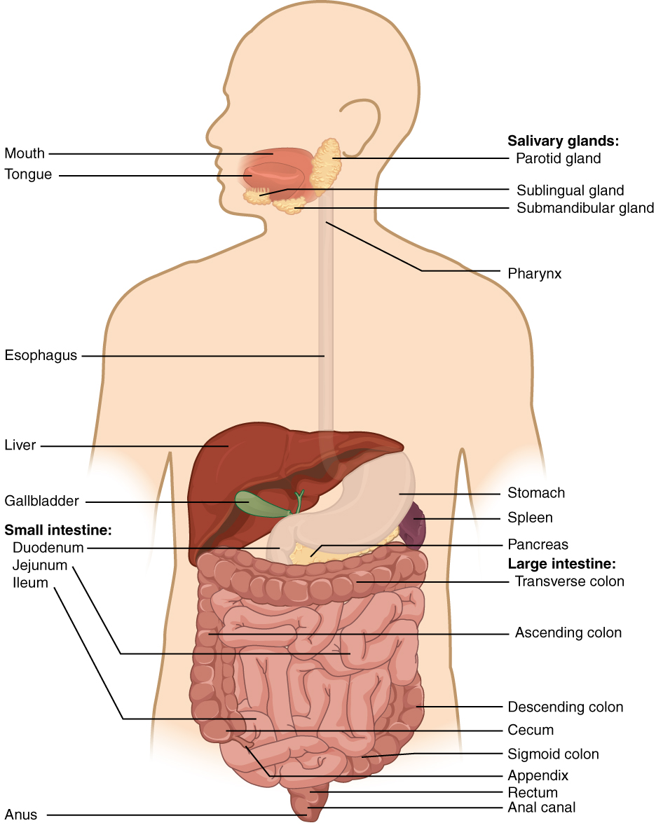
Figure 4. All digestive organs play integral roles in the life-sustaining process of digestion.
The first step in the digestive system can actually begin before the food is even in your mouth. When you smell or see something that you just have to eat, you start to salivate in anticipation of eating, thus beginning the digestive process.
Food is the body’s source of fuel. Nutrients in food give the body’s cells the energy they need to operate. Before food can be used it has to be broken down into tiny little pieces so it can be absorbed and used by the body. In humans, proteins need to be broken down into amino acids, starches into sugars, and fats into fatty acids and glycerol.
During digestion two main processes occur at the same time:
- Mechanical Digestion: larger pieces of food get broken down into smaller pieces while being prepared for chemical digestion. Mechanical digestion starts in the mouth and continues in to the stomach.
- Chemical Digestion: several different enzymes break down macromolecules into smaller molecules that can be more efficiently absorbed. Chemical digestion starts with saliva and continues into the intestines. The major enzymes involved in chemical digestion are shown in the table below.
The digestive system is made up by the alimentary canal, or the digestive tract, and other abdominal organs that play a part in digestion such as the liver and the pancreas. The alimentary canal is the long tube of organs that runs from the mouth (where the food enters) to the anus (where indigestible waste leaves). The organs in the alimentary canal include the mouth( for mastication),esophagus, stomach and the intestines. The average adult digestive tract is about thirty feet (30′) long. While in the digestive tract the food is really passing through the body rather than being in the body. The smooth muscles of the tubular digestive organs move the food efficiently along as it is broken down into absorb-able atoms and molecules. During absorption, the nutrients that come from food (such as proteins, fats, carbohydrates, vitamins, and minerals) pass through the wall of the small intestine and into the bloodstream and lymph. In this way nutrients can be distributed throughout the rest of the body. In the large intestine there is reabsorption of water and absorption of some minerals as feces are formed. The parts of the food that the body passes out through the anus is known as feces.
| Enzyme | Produced In | Site of Release | pH Level |
|---|---|---|---|
| Carbohydrate Digestion | |||
| Salivary amylase | Salivary glands | Mouth | Neutral |
| Pancreatic amylase | Pancreas | Small intestine | Basic |
| Maltase | Small intestine | Small intestine | Basic |
| Protein Digestion | |||
| Pepsin | Gastric glands | Stomach | Acidic |
| Trypsin | Pancreas | Small intestine | Basic |
| Peptidases | Small intestine | Small intestine | Basic |
| Nucleic Acid Digestion | |||
| Nuclease | Pancreas | Small intestine | Basic |
| Nucleosidases | Pancreas | Small intestine | Basic |
| Fat Digestion | |||
| Lipase | Pancreas | Small intestine | Basic |
Video Review
Watch this video series introducing the digestive system:
Check Your Understanding
Answer the question(s) below to see how well you understand the topics covered in the previous section. This short quiz does not count toward your grade in the class, and you can retake it an unlimited number of times.
Use this quiz to check your understanding and decide whether to (1) study the previous section further or (2) move on to the next section.
Candela Citations
- Introduction to Cell Maintenance Systems. Authored by: Shelli Carter and Lumen Learning. Provided by: Lumen Learning. License: CC BY: Attribution
- Human Physiology. Provided by: Wikibooks. Located at: https://en.wikibooks.org/wiki/Human_Physiology. License: CC BY-SA: Attribution-ShareAlike
- Heart Illustration. Provided by: OpenStax. Located at: http://cnx.org/contents/185cbf87-c72e-48f5-b51e-f14f21b5eabd@10.8. Project: Biology. License: CC BY: Attribution. License Terms: Download for free at http://cnx.org/contents/185cbf87-c72e-48f5-b51e-f14f21b5eabd@10.8
- Respiratory system 1- Functions. Authored by: Wendy Riggs. Located at: https://youtu.be/FmbhxVRUJZI. License: CC BY: Attribution
- An eText of Human Anatomy and Physiology. Authored by: Dr. Bruce Forciea. Located at: http://www.bruceforciea.com/etextchapters/etexthumananatrevmay12.pdf. License: CC BY: Attribution
- Respiratory tract. Provided by: OpenStax. Located at: http://cnx.org/contents/14fb4ad7-39a1-4eee-ab6e-3ef2482e3e22@8.79. Project: Anatomy & Physiology. License: CC BY: Attribution. License Terms: Download for free at http://cnx.org/contents/14fb4ad7-39a1-4eee-ab6e-3ef2482e3e22@8.79
- Anatomy 04- Digestive System. Authored by: Wendy Riggs. Located at: https://youtu.be/iYh1B7s8h28?list=PL5GRRRmaGVqVCPc8dCRfTMHfPioZK5W_0. License: CC BY: Attribution
- Digestive System. Provided by: OpenStax. Located at: http://cnx.org/contents/14fb4ad7-39a1-4eee-ab6e-3ef2482e3e22@8.80. Project: Anatomy & Physiology. License: CC BY: Attribution. License Terms: Download for free at http://cnx.org/contents/14fb4ad7-39a1-4eee-ab6e-3ef2482e3e22@8.80
- The Circulatory System. Authored by: Bozeman Science. Located at: https://youtu.be/NJzJKvkWWDc. License: All Rights Reserved. License Terms: Standard YouTube License
- The Immune System. Authored by: Bozeman Science. Located at: https://youtu.be/z3M0vU3Dv8E. License: All Rights Reserved. License Terms: Standard YouTube License