Identify basic common structures of plants
While individual plant species are unique, all share a common structure: a plant body consisting of stems, roots, and leaves. They all transport water, minerals, and sugars produced through photosynthesis through the plant body in a similar manner. All plant species also respond to environmental factors, such as light, gravity, competition, temperature, and predation.
Learning Objectives
- Discuss features of plant cells
- Identify the different tissue types and organ systems in plants
- Describe the main function and basic structure of stems
- Identify the structure and function of a typical leaf
- Identify the two types of root systems
Plant Cells

Figure 1. A section of a pine embryo.
Why do plant cells look like little rectangles? Look at Figure 1 and notice how all the cells seem to stack on each other, with no spaces in between. Might this allow the cells to form structures that can grow upright?
Organs in Plants?
Your body includes organ systems, such as the digestive system, made of individual organs, such as the stomach, liver, and pancreas, which work together to carry out a certain function (in this case, breaking down and absorbing food). These organs, in turn, are made of different kinds of tissues, which are groups of cells which work together to perform a specific job. For example, your stomach is made of muscle tissue to facilitate movement and glandular tissue to secrete enzymes for chemical breakdown of food molecules. These tissues, in turn, are made of cells specialized in shape, size, and component organelles, such as mitochondria for energy and microtubules for movement.
Plants, too, are made of organs, which in turn are made of tissues. Plant tissues, like ours, are constructed of specialized cells, which in turn contain specific organelles. It is these cells, tissues, and organs that carry out the dramatic lives of plants.
Plant Cells
Plant cells resemble other eukaryotic cells in many ways. For example, they are enclosed by a plasma membrane and have a nucleus and other membrane-bound organelles. A typical plant cell is represented by the diagram in Figure 2.
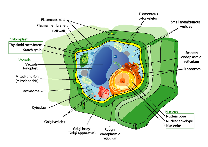
Figure 2. Plant cells have all the same structures as animal cells, plus some additional structures. Can you identify the unique plant structures in the diagram?
Plant Cell Structures
Structures found in plant cells but not animal cells include a large central vacuole, cell wall, and plastids such as chloroplasts.
- The large central vacuole is surrounded by its own membrane and contains water and dissolved substances. Its primary role is to maintain pressure against the inside of the cell wall, giving the cell shape and helping to support the plant.
- The cell wall is located outside the cell membrane. It consists mainly of cellulose and may also contain lignin , which makes it more rigid. The cell wall shapes, supports, and protects the cell. It prevents the cell from absorbing too much water and bursting. It also keeps large, damaging molecules out of the cell.
- Plastids are membrane-bound organelles with their own DNA. Examples are chloroplasts and chromoplasts. Chloroplasts contain the green pigment chlorophyll and carry out photosynthesis. Chromoplasts make and store other pigments. They give flower petals their bright colors.
Types of Plant Cells
There are three basic types of cells in most plants. These cells make up ground tissue, which will be discussed in another concept. The three types of cells are described in table below. The different types of plant cells have different structures and functions.
| Type of Cell | Structure | Functions | Example |
|---|---|---|---|
| Parenchymal | cube-shaped
loosely packed thin-walled relatively unspecialized contain chloroplasts |
photosynthesis
cellular respiration storage |
food storage tissues of potatoes
|
| Collenchymal | elongated
irregularly thickened walls |
support
wind resistance |
strings running through a stalk of celery
|
| Sclerenchymal | very thick cell walls containing lignin | support
strength |
tough fibers in jute (used to make rope)
|


Plant Tissues
Plants are multicellular eukaryotes with tissue systems made of various cell types that carry out specific functions. Plant tissue systems fall into one of two general types: meristematic tissue and permanent (or non-meristematic) tissue. Cells of the meristematic tissue are found in meristems, which are plant regions of continuous cell division and growth. Meristematic tissue cells are either undifferentiated or incompletely differentiated, and they continue to divide and contribute to the growth of the plant. In contrast, permanent tissue consists of plant cells that are no longer actively dividing.
Meristematic tissues consist of three types, based on their location in the plant. Apical meristems contain meristematic tissue located at the tips of stems and roots, which enable a plant to extend in length. Lateral meristems facilitate growth in thickness or girth in a maturing plant. Intercalary meristems occur only in monocots, at the bases of leaf blades and at nodes (the areas where leaves attach to a stem). This tissue enables the monocot leaf blade to increase in length from the leaf base; for example, it allows lawn grass leaves to elongate even after repeated mowing.
Meristems produce cells that quickly differentiate, or specialize, and become permanent tissue. Such cells take on specific roles and lose their ability to divide further. They differentiate into three main types: dermal, vascular, and ground tissue. Dermal tissue covers and protects the plant, and vascular tissue transports water, minerals, and sugars to different parts of the plant. Ground tissue serves as a site for photosynthesis, provides a supporting matrix for the vascular tissue, and helps to store water and sugars.

Figure 3. This light micrograph shows a cross section of a squash (Curcurbita maxima) stem. Each teardrop-shaped vascular bundle consists of large xylem vessels toward the inside and smaller phloem cells toward the outside. Xylem cells, which transport water and nutrients from the roots to the rest of the plant, are dead at functional maturity. Phloem cells, which transport sugars and other organic compounds from photosynthetic tissue to the rest of the plant, are living. The vascular bundles are encased in ground tissue and surrounded by dermal tissue. (credit: modification of work by “(biophotos)”/Flickr; scale-bar data from Matt Russell)
Secondary tissues are either simple (composed of similar cell types) or complex (composed of different cell types). Dermal tissue, for example, is a simple tissue that covers the outer surface of the plant and controls gas exchange. Vascular tissue is an example of a complex tissue, and is made of two specialized conducting tissues: xylem and phloem. Xylem tissue transports water and nutrients from the roots to different parts of the plant, and includes three different cell types: vessel elements and tracheids (both of which conduct water), and xylem parenchyma. Phloem tissue, which transports organic compounds from the site of photosynthesis to other parts of the plant, consists of four different cell types: sieve cells (which conduct photosynthates), companion cells, phloem parenchyma, and phloem fibers. Unlike xylem conducting cells, phloem conducting cells are alive at maturity. The xylem and phloem always lie adjacent to each other (Figure 3). In stems, the xylem and the phloem form a structure called a vascular bundle; in roots, this is termed the vascular stele or vascular cylinder.
All animals are made of four types of tissue: epidermal, muscle, nerve, and connective tissues. Plants, too, are built of tissues, but not surprisingly, their very different lifestyles derive from different kinds of tissues. All three types of plant cells are found in most plant tissues. Three major types of plant tissues are dermal, ground, and vascular tissues.
Dermal Tissue
The dermal tissue of the stem consists primarily of epidermis, a single layer of cells covering and protecting the underlying tissue. Woody plants have a tough, waterproof outer layer of cork cells commonly known as bark, which further protects the plant from damage. Epidermal cells are the most numerous and least differentiated of the cells in the epidermis. The epidermis of a leaf also contains openings known as stomata, through which the exchange of gases takes place (Figure 4). Two cells, known as guard cells, surround each leaf stoma, controlling its opening and closing and thus regulating the uptake of carbon dioxide and the release of oxygen and water vapor. Trichomes are hair-like structures on the epidermal surface. They help to reduce transpiration (the loss of water by aboveground plant parts), increase solar reflectance, and store compounds that defend the leaves against predation by herbivores.

Figure 4. Openings called stomata (singular: stoma) allow a plant to take up carbon dioxide and release oxygen and water vapor. The (a) colorized scanning-electron micrograph shows a closed stoma of a dicot. Each stoma is flanked by two guard cells that regulate its (b) opening and closing. The (c) guard cells sit within the layer of epidermal cells (credit a: modification of work by Louisa Howard, Rippel Electron Microscope Facility, Dartmouth College; credit b: modification of work by June Kwak, University of Maryland; scale-bar data from Matt Russell)
Vascular Tissue
The xylem and phloem that make up the vascular tissue of the stem are arranged in distinct strands called vascular bundles, which run up and down the length of the stem. When the stem is viewed in cross section, the vascular bundles of dicot stems are arranged in a ring. In plants with stems that live for more than one year, the individual bundles grow together and produce the characteristic growth rings. In monocot stems, the vascular bundles are randomly scattered throughout the ground tissue (Figure 5).

Figure 5. In (a) dicot stems, vascular bundles are arranged around the periphery of the ground tissue. The xylem tissue is located toward the interior of the vascular bundle, and phloem is located toward the exterior. Sclerenchyma fibers cap the vascular bundles. In (b) monocot stems, vascular bundles composed of xylem and phloem tissues are scattered throughout the ground tissue.
Xylem tissue has three types of cells: xylem parenchyma, tracheids, and vessel elements. The latter two types conduct water and are dead at maturity. Tracheids are xylem cells with thick secondary cell walls that are lignified. Water moves from one tracheid to another through regions on the side walls known as pits, where secondary walls are absent. Vessel elements are xylem cells with thinner walls; they are shorter than tracheids. Each vessel element is connected to the next by means of a perforation plate at the end walls of the element. Water moves through the perforation plates to travel up the plant.
Phloem tissue is composed of sieve-tube cells, companion cells, phloem parenchyma, and phloem fibers. A series of sieve-tube cells (also called sieve-tube elements) are arranged end to end to make up a long sieve tube, which transports organic substances such as sugars and amino acids. The sugars flow from one sieve-tube cell to the next through perforated sieve plates, which are found at the end junctions between two cells. Although still alive at maturity, the nucleus and other cell components of the sieve-tube cells have disintegrated. Companion cells are found alongside the sieve-tube cells, providing them with metabolic support. The companion cells contain more ribosomes and mitochondria than the sieve-tube cells, which lack some cellular organelles.
Ground Tissue
Ground tissue is mostly made up of parenchyma cells, but may also contain collenchyma and sclerenchyma cells that help support the stem. The ground tissue towards the interior of the vascular tissue in a stem or root is known as pith, while the layer of tissue between the vascular tissue and the epidermis is known as the cortex.
Plant Organs
Like animals, plants contain cells with organelles in which specific metabolic activities take place. Unlike animals, however, plants use energy from sunlight to form sugars during photosynthesis. In addition, plant cells have cell walls, plastids, and a large central vacuole: structures that are not found in animal cells. Each of these cellular structures plays a specific role in plant structure and function.
In plants, just as in animals, similar cells working together form a tissue. When different types of tissues work together to perform a unique function, they form an organ; organs working together form organ systems. Vascular plants have two distinct organ systems: a shoot system, and a root system. The shoot system consists of two portions: the vegetative (non-reproductive) parts of the plant, such as the leaves and the stems, and the reproductive parts of the plant, which include flowers and fruits. The shoot system generally grows above ground, where it absorbs the light needed for photosynthesis. The root system, which supports the plants and absorbs water and minerals, is usually underground. Figure 6 shows the organ systems of a typical plant.

Figure 6. The shoot system of a plant consists of leaves, stems, flowers, and fruits. The root system anchors the plant while absorbing water and minerals from the soil.
Stems

Figure 7. Leaves are attached to the plant stem at areas called nodes. An internode is the stem region between two nodes. The petiole is the stalk connecting the leaf to the stem. The leaves just above the nodes arose from axillary buds.
Stems are a part of the shoot system of a plant. They may range in length from a few millimeters to hundreds of meters, and also vary in diameter, depending on the plant type. Stems are usually above ground, although the stems of some plants, such as the potato, also grow underground. Stems may be herbaceous (soft) or woody in nature. Their main function is to provide support to the plant, holding leaves, flowers and buds; in some cases, stems also store food for the plant. A stem may be unbranched, like that of a palm tree, or it may be highly branched, like that of a magnolia tree. The stem of the plant connects the roots to the leaves, helping to transport absorbed water and minerals to different parts of the plant. It also helps to transport the products of photosynthesis, namely sugars, from the leaves to the rest of the plant.
Plant stems, whether above or below ground, are characterized by the presence of nodes and internodes (Figure 7). Nodes are points of attachment for leaves, aerial roots, and flowers. The stem region between two nodes is called an internode. The stalk that extends from the stem to the base of the leaf is the petiole. An axillary bud is usually found in the axil—the area between the base of a leaf and the stem—where it can give rise to a branch or a flower. The apex (tip) of the shoot contains the apical meristem within the apical bud.
Stem Anatomy

Figure 8. The stem of common St John’s Wort (Hypericum perforatum) is shown in cross section in this light micrograph. (credit: Rolf-Dieter Mueller)
The stem and other plant organs arise from the ground tissue, and are primarily made up of simple tissues formed from three types of cells: parenchyma, collenchyma, and sclerenchyma cells.
Parenchyma cells are the most common plant cells (Figure 8). They are found in the stem, the root, the inside of the leaf, and the pulp of the fruit. Parenchyma cells are responsible for metabolic functions, such as photosynthesis, and they help repair and heal wounds. Some parenchyma cells also store starch. In Figure 8, we see the central pith (greenish-blue, in the center) and peripheral cortex (narrow zone 3–5 cells thick just inside the epidermis); both are composed of parenchyma cells. Vascular tissue composed of xylem (red) and phloem tissue (green, between the xylem and cortex) surrounds the pith.
Collenchyma cells are elongated cells with unevenly thickened walls (Figure 9). They provide structural support, mainly to the stem and leaves. These cells are alive at maturity and are usually found below the epidermis. The “strings” of a celery stalk are an example of collenchyma cells.

Figure 9. Collenchyma cell walls are uneven in thickness, as seen in this light micrograph. They provide support to plant structures. (credit: modification of work by Carl Szczerski; scale-bar data from Matt Russell)
Sclerenchyma cells also provide support to the plant, but unlike collenchyma cells, many of them are dead at maturity. There are two types of sclerenchyma cells: fibers and sclereids. Both types have secondary cell walls that are thickened with deposits of lignin, an organic compound that is a key component of wood. Fibers are long, slender cells; sclereids are smaller-sized. Sclereids give pears their gritty texture. Humans use sclerenchyma fibers to make linen and rope (Figure 10).

Figure 10. The central pith and outer cortex of the (a) flax stem are made up of parenchyma cells. Inside the cortex is a layer of sclerenchyma cells, which make up the fibers in flax rope and clothing. Humans have grown and harvested flax for thousands of years. In (b) this drawing, fourteenth-century women prepare linen. The (c) flax plant is grown and harvested for its fibers, which are used to weave linen, and for its seeds, which are the source of linseed oil. (credit a: modification of work by Emmanuel Boutet based on original work by Ryan R. MacKenzie; credit c: modification of work by Brian Dearth; scale-bar data from Matt Russell)
Practice Question
Which layers of the stem are made of parenchyma cells?
- cortex and pith
- phloem
- sclerenchyma
- xylem
Stem Modifications
Some plant species have modified stems that are especially suited to a particular habitat and environment (Figure 11). A rhizome is a modified stem that grows horizontally underground and has nodes and internodes. Vertical shoots may arise from the buds on the rhizome of some plants, such as ginger and ferns. Corms are similar to rhizomes, except they are more rounded and fleshy (such as in gladiolus). Corms contain stored food that enables some plants to survive the winter. Stolons are stems that run almost parallel to the ground, or just below the surface, and can give rise to new plants at the nodes. Runners are a type of stolon that runs above the ground and produces new clone plants at nodes at varying intervals: strawberries are an example. Tubers are modified stems that may store starch, as seen in the potato (Solanum sp.). Tubers arise as swollen ends of stolons, and contain many adventitious or unusual buds (familiar to us as the “eyes” on potatoes). A bulb, which functions as an underground storage unit, is a modification of a stem that has the appearance of enlarged fleshy leaves emerging from the stem or surrounding the base of the stem, as seen in the iris.

Figure 11. Stem modifications enable plants to thrive in a variety of environments. Shown are (a) ginger (Zingiber officinale) rhizomes, (b) a carrion flower (Amorphophallus titanum) corm (c) Rhodes grass (Chloris gayana) stolons, (d) strawberry (Fragaria ananassa) runners, (e) potato (Solanum tuberosum) tubers, and (f) red onion (Allium) bulbs. (credit a: modification of work by Maja Dumat; credit c: modification of work by Harry Rose; credit d: modification of work by Rebecca Siegel; credit e: modification of work by Scott Bauer, USDA ARS; credit f: modification of work by Stephen Ausmus, USDA ARS)
Watch botanist Wendy Hodgson, of Desert Botanical Garden in Phoenix, Arizona, explain how agave plants were cultivated for food hundreds of years ago in the Arizona desert in this video: Finding the Roots of an Ancient Crop.
Some aerial modifications of stems are tendrils and thorns (Figure 12). Tendrils are slender, twining strands that enable a plant (like a vine or pumpkin) to seek support by climbing on other surfaces. Thorns are modified branches appearing as sharp outgrowths that protect the plant; common examples include roses, Osage orange and devil’s walking stick.

Figure 12. Found in southeastern United States, (a) buckwheat vine (Brunnichia ovata) is a weedy plant that climbs with the aid of tendrils. This one is shown climbing up a wooden stake. (b) Thorns are modified branches. (credit a: modification of work by Christopher Meloche, USDA ARS; credit b: modification of work by “macrophile”/Flickr)
Leaves
Leaves are the main sites for photosynthesis: the process by which plants synthesize food. Most leaves are usually green, due to the presence of chlorophyll in the leaf cells. However, some leaves may have different colors, caused by other plant pigments that mask the green chlorophyll.
The thickness, shape, and size of leaves are adapted to the environment. Each variation helps a plant species maximize its chances of survival in a particular habitat. Usually, the leaves of plants growing in tropical rainforests have larger surface areas than those of plants growing in deserts or very cold conditions, which are likely to have a smaller surface area to minimize water loss.
Structure of a Typical Leaf

Figure 13. Deceptively simple in appearance, a leaf is a highly efficient structure.
Each leaf typically has a leaf blade called the lamina, which is also the widest part of the leaf. Some leaves are attached to the plant stem by a petiole. Leaves that do not have a petiole and are directly attached to the plant stem are called sessile leaves. Small green appendages usually found at the base of the petiole are known as stipules. Most leaves have a midrib, which travels the length of the leaf and branches to each side to produce veins of vascular tissue. The edge of the leaf is called the margin. Figure 13 shows the structure of a typical eudicot leaf.
Within each leaf, the vascular tissue forms veins. The arrangement of veins in a leaf is called the venation pattern. Monocots and dicots differ in their patterns of venation (Figure 14). Monocots have parallel venation; the veins run in straight lines across the length of the leaf without converging at a point. In dicots, however, the veins of the leaf have a net-like appearance, forming a pattern known as reticulate venation. One extant plant, the Ginkgo biloba, has dichotomous venation where the veins fork.

Figure 14. (a) Tulip (Tulipa), a monocot, has leaves with parallel venation. The netlike venation in this (b) linden (Tilia cordata) leaf distinguishes it as a dicot. The (c) Ginkgo biloba tree has dichotomous venation. (credit a photo: modification of work by “Drewboy64”/Wikimedia Commons; credit b photo: modification of work by Roger Griffith; credit c photo: modification of work by “geishaboy500″/Flickr; credit abc illustrations: modification of work by Agnieszka Kwiecień)
Leaf Arrangement
The arrangement of leaves on a stem is known as phyllotaxy. The number and placement of a plant’s leaves will vary depending on the species, with each species exhibiting a characteristic leaf arrangement. Leaves are classified as either alternate, spiral, or opposite. Plants that have only one leaf per node have leaves that are said to be either alternate—meaning the leaves alternate on each side of the stem in a flat plane—or spiral, meaning the leaves are arrayed in a spiral along the stem. In an opposite leaf arrangement, two leaves arise at the same point, with the leaves connecting opposite each other along the branch. If there are three or more leaves connected at a node, the leaf arrangement is classified as whorled.
Leaf Form
Leaves may be simple or compound (Figure 15). In a simple leaf, the blade is either completely undivided—as in the banana leaf—or it has lobes, but the separation does not reach the midrib, as in the maple leaf. In a compound leaf, the leaf blade is completely divided, forming leaflets, as in the locust tree. Each leaflet may have its own stalk, but is attached to the rachis. A palmately compound leaf resembles the palm of a hand, with leaflets radiating outwards from one point Examples include the leaves of poison ivy, the buckeye tree, or the familiar houseplant Schefflera sp. (common name “umbrella plant”). Pinnately compound leaves take their name from their feather-like appearance; the leaflets are arranged along the midrib, as in rose leaves (Rosa sp.), or the leaves of hickory, pecan, ash, or walnut trees.

Figure 15. Leaves may be simple or compound. In simple leaves, the lamina is continuous. The (a) banana plant (Musa sp.) has simple leaves. In compound leaves, the lamina is separated into leaflets. Compound leaves may be palmate or pinnate. In (b) palmately compound leaves, such as those of the horse chestnut (Aesculus hippocastanum), the leaflets branch from the petiole. In (c) pinnately compound leaves, the leaflets branch from the midrib, as on a scrub hickory (Carya floridana). The (d) honey locust has double compound leaves, in which leaflets branch from the veins. (credit a: modification of work by “BazzaDaRambler”/Flickr; credit b: modification of work by Roberto Verzo; credit c: modification of work by Eric Dion; credit d: modification of work by Valerie Lykes)
Leaf Structure and Function
The outermost layer of the leaf is the epidermis; it is present on both sides of the leaf and is called the upper and lower epidermis, respectively. Botanists call the upper side the adaxial surface (or adaxis) and the lower side the abaxial surface (or abaxis). The epidermis helps in the regulation of gas exchange. It contains stomata (Figure 16): openings through which the exchange of gases takes place. Two guard cells surround each stoma, regulating its opening and closing.

Figure 16. Visualized at 500x with a scanning electron microscope, several stomata are clearly visible on (a) the surface of this sumac (Rhus glabra) leaf. At 5,000x magnification, the guard cells of (b) a single stoma from lyre-leaved sand cress (Arabidopsis lyrata) have the appearance of lips that surround the opening. In this (c) light micrograph cross-section of an A. lyrata leaf, the guard cell pair is visible along with the large, sub-stomatal air space in the leaf. (credit: modification of work by Robert R. Wise; part c scale-bar data from Matt Russell)
The epidermis is usually one cell layer thick; however, in plants that grow in very hot or very cold conditions, the epidermis may be several layers thick to protect against excessive water loss from transpiration. A waxy layer known as the cuticle covers the leaves of all plant species. The cuticle reduces the rate of water loss from the leaf surface. Other leaves may have small hairs (trichomes) on the leaf surface. Trichomes help to deter herbivory by restricting insect movements, or by storing toxic or bad-tasting compounds; they can also reduce the rate of transpiration by blocking air flow across the leaf surface (Figure 17).

Figure 17. Trichomes give leaves a fuzzy appearance as in this (a) sundew (Drosera sp.). Leaf trichomes include (b) branched trichomes on the leaf of Arabidopsis lyrata and (c) multibranched trichomes on a mature Quercus marilandica leaf. (credit a: John Freeland; credit b, c: modification of work by Robert R. Wise; scale-bar data from Matt Russell)
Below the epidermis of dicot leaves are layers of cells known as the mesophyll, or “middle leaf.” The mesophyll of most leaves typically contains two arrangements of parenchyma cells: the palisade parenchyma and spongy parenchyma (Figure 18). The palisade parenchyma (also called the palisade mesophyll) has column-shaped, tightly packed cells, and may be present in one, two, or three layers. Below the palisade parenchyma are loosely arranged cells of an irregular shape. These are the cells of the spongy parenchyma (or spongy mesophyll). The air space found between the spongy parenchyma cells allows gaseous exchange between the leaf and the outside atmosphere through the stomata. In aquatic plants, the intercellular spaces in the spongy parenchyma help the leaf float. Both layers of the mesophyll contain many chloroplasts. Guard cells are the only epidermal cells to contain chloroplasts.
In the leaf drawing (Figure 18a), the central mesophyll is sandwiched between an upper and lower epidermis. The mesophyll has two layers: an upper palisade layer comprised of tightly packed, columnar cells, and a lower spongy layer, comprised of loosely packed, irregularly shaped cells. Stomata on the leaf underside allow gas exchange. A waxy cuticle covers all aerial surfaces of land plants to minimize water loss. These leaf layers are clearly visible in the scanning electron micrograph (Figure 18b). The numerous small bumps in the palisade parenchyma cells are chloroplasts. Chloroplasts are also present in the spongy parenchyma, but are not as obvious. The bumps protruding from the lower surface of the leave are glandular trichomes, which differ in structure from the stalked trichomes in Figure 17.
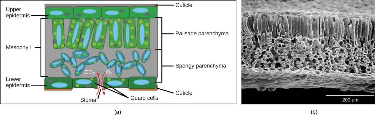
Figure 18. (a) Leaf drawing (b) Scanning electron micrograph of a leaf. (credit b: modification of work by Robert R. Wise)

Figure 19. This scanning electron micrograph shows xylem and phloem in the leaf vascular bundle from the lyre-leaved sand cress (Arabidopsis lyrata). (credit: modification of work by Robert R. Wise; scale-bar data from Matt Russell)
Like the stem, the leaf contains vascular bundles composed of xylem and phloem (Figure 19). The xylem consists of tracheids and vessels, which transport water and minerals to the leaves. The phloem transports the photosynthetic products from the leaf to the other parts of the plant. A single vascular bundle, no matter how large or small, always contains both xylem and phloem tissues.
Leaf Adaptations
Coniferous plant species that thrive in cold environments, like spruce, fir, and pine, have leaves that are reduced in size and needle-like in appearance. These needle-like leaves have sunken stomata and a smaller surface area: two attributes that aid in reducing water loss. In hot climates, plants such as cacti have leaves that are reduced to spines, which in combination with their succulent stems, help to conserve water. Many aquatic plants have leaves with wide lamina that can float on the surface of the water, and a thick waxy cuticle on the leaf surface that repels water.
Watch “The Pale Pitcher Plant” episode of the video series Plants Are Cool, Too, a Botanical Society of America video about a carnivorous plant species found in Louisiana.
In Summary: Leaves
Leaves are the main site of photosynthesis. A typical leaf consists of a lamina (the broad part of the leaf, also called the blade) and a petiole (the stalk that attaches the leaf to a stem). The arrangement of leaves on a stem, known as phyllotaxy, enables maximum exposure to sunlight. Each plant species has a characteristic leaf arrangement and form. The pattern of leaf arrangement may be alternate, opposite, or spiral, while leaf form may be simple or compound. Leaf tissue consists of the epidermis, which forms the outermost cell layer, and mesophyll and vascular tissue, which make up the inner portion of the leaf. In some plant species, leaf form is modified to form structures such as tendrils, spines, bud scales, and needles.
Roots
The roots of seed plants have three major functions: anchoring the plant to the soil, absorbing water and minerals and transporting them upwards, and storing the products of photosynthesis. Some roots are modified to absorb moisture and exchange gases. Most roots are underground. Some plants, however, also have adventitious roots, which emerge above the ground from the shoot.
Types of Root Systems
Root systems are mainly of two types (Figure 20). Dicots have a tap root system, while monocots have a fibrous root system. A tap root system has a main root that grows down vertically, and from which many smaller lateral roots arise. Dandelions are a good example; their tap roots usually break off when trying to pull these weeds, and they can regrow another shoot from the remaining root). A tap root system penetrates deep into the soil. In contrast, a fibrous root system is located closer to the soil surface, and forms a dense network of roots that also helps prevent soil erosion (lawn grasses are a good example, as are wheat, rice, and corn). Some plants have a combination of tap roots and fibrous roots. Plants that grow in dry areas often have deep root systems, whereas plants growing in areas with abundant water are likely to have shallower root systems.

Figure 20. (a) Tap root systems have a main root that grows down, while (b) fibrous root systems consist of many small roots. (credit b: modification of work by “Austen Squarepants”/Flickr)
Root Growth and Anatomy

Figure 21. A longitudinal view of the root reveals the zones of cell division, elongation, and maturation. Cell division occurs in the apical meristem.
Root growth begins with seed germination. When the plant embryo emerges from the seed, the radicle of the embryo forms the root system. The tip of the root is protected by the root cap, a structure exclusive to roots and unlike any other plant structure. The root cap is continuously replaced because it gets damaged easily as the root pushes through soil. The root tip can be divided into three zones: a zone of cell division, a zone of elongation, and a zone of maturation and differentiation (Figure 21). The zone of cell division is closest to the root tip; it is made up of the actively dividing cells of the root meristem. The zone of elongation is where the newly formed cells increase in length, thereby lengthening the root. Beginning at the first root hair is the zone of cell maturation where the root cells begin to differentiate into special cell types. All three zones are in the first centimeter or so of the root tip.
The root has an outer layer of cells called the epidermis, which surrounds areas of ground tissue and vascular tissue. The epidermis provides protection and helps in absorption. Root hairs, which are extensions of root epidermal cells, increase the surface area of the root, greatly contributing to the absorption of water and minerals.

Figure 22. Staining reveals different cell types in this light micrograph of a wheat (Triticum) root cross section. Sclerenchyma cells of the exodermis and xylem cells stain red, and phloem cells stain blue. Other cell types stain black. The stele, or vascular tissue, is the area inside endodermis (indicated by a green ring). Root hairs are visible outside the epidermis. (credit: scale-bar data from Matt Russell)
Inside the root, the ground tissue forms two regions: the cortex and the pith (Figure 22). Compared to stems, roots have lots of cortex and little pith. Both regions include cells that store photosynthetic products. The cortex is between the epidermis and the vascular tissue, whereas the pith lies between the vascular tissue and the center of the root.
The vascular tissue in the root is arranged in the inner portion of the root, which is called the stele (Figure 23). A layer of cells known as the endodermis separates the stele from the ground tissue in the outer portion of the root. The endodermis is exclusive to roots, and serves as a checkpoint for materials entering the root’s vascular system. A waxy substance called suberin is present on the walls of the endodermal cells. This waxy region, known as the Casparian strip, forces water and solutes to cross the plasma membranes of endodermal cells instead of slipping between the cells. This ensures that only materials required by the root pass through the endodermis, while toxic substances and pathogens are generally excluded. The outermost cell layer of the root’s vascular tissue is the pericycle, an area that can give rise to lateral roots. In dicot roots, the xylem and phloem of the stele are arranged alternately in an X shape, whereas in monocot roots, the vascular tissue is arranged in a ring around the pith.

Figure 23. In (left) typical dicots, the vascular tissue forms an X shape in the center of the root. In (right) typical monocots, the phloem cells and the larger xylem cells form a characteristic ring around the central pith.
Root Modifications

Figure 24. Many vegetables are modified roots.
Root structures may be modified for specific purposes. For example, some roots are bulbous and store starch. Aerial roots and prop roots are two forms of aboveground roots that provide additional support to anchor the plant. Tap roots, such as carrots, turnips, and beets, are examples of roots that are modified for food storage (Figure 24).
Epiphytic roots enable a plant to grow on another plant. For example, the epiphytic roots of orchids develop a spongy tissue to absorb moisture. The banyan tree (Ficus sp.) begins as an epiphyte, germinating in the branches of a host tree; aerial roots develop from the branches and eventually reach the ground, providing additional support (Figure 25). In screwpine (Pandanus sp.), a palm-like tree that grows in sandy tropical soils, aboveground prop roots develop from the nodes to provide additional support.

Figure 25. The (a) banyan tree, also known as the strangler fig, begins life as an epiphyte in a host tree. Aerial roots extend to the ground and support the growing plant, which eventually strangles the host tree. The (b) screwpine develops aboveground roots that help support the plant in sandy soils. (credit a: modification of work by “psyberartist”/Flickr; credit b: modification of work by David Eikhoff)
Practice Questions
Compare a tap root system with a fibrous root system. For each type, name a plant that provides a food in the human diet. Which type of root system is found in monocots? Which type of root system is found in dicots?
What might happen to a root if the pericycle disappeared?
Check Your Understanding
Answer the question(s) below to see how well you understand the topics covered in the previous section. This short quiz does not count toward your grade in the class, and you can retake it an unlimited number of times.
Use this quiz to check your understanding and decide whether to (1) study the previous section further or (2) move on to the next section.
Candela Citations
- Introduction to Plant Structures. Authored by: Shelli Carter and Lumen Learning. Provided by: Lumen Learning. License: CC BY: Attribution
- Biology. Provided by: OpenStax CNX. Located at: http://cnx.org/contents/185cbf87-c72e-48f5-b51e-f14f21b5eabd@10.8. License: CC BY: Attribution. License Terms: Download for free at http://cnx.org/contents/185cbf87-c72e-48f5-b51e-f14f21b5eabd@10.8
- Plant Cells. Authored by: Douglas Wilkin, Ph.D. and Jean Brainard, Ph.D.. Provided by: CK-12. Located at: http://www.ck12.org/biology/Plant-Cells/lesson/Plant-Cells/r34/. License: CC BY-NC: Attribution-NonCommercial
- Plant Tissues. Authored by: Douglas Wilkin, Ph.D. and Jean Brainard, Ph.D.. Provided by: CK-12. Located at: http://www.ck12.org/biology/Plant-Tissues/lesson/Plant-Tissues/r34/. License: CC BY-NC: Attribution-NonCommercial
- Finding The Roots Of An Ancient Crop. Authored by: SciFri. Located at: https://youtu.be/0AXF9RmqBtg. License: All Rights Reserved. License Terms: Standard YouTube License
- Plants Are Cool Too! The Pale Pitcher Plant (Episode 1 - Sarracenia alata. Authored by: BotanicalSociety. Located at: https://youtu.be/uak3m_q-HDo. License: All Rights Reserved. License Terms: Standard YouTube License
- Life - Venus Flytraps: Jaws of Death - BBC One. Authored by: BBC. Located at: https://youtu.be/O7eQKSf0LmY. License: All Rights Reserved. License Terms: Standard YouTube License