Learning Objectives
- Describe the functions of different major areas of the brain and endocrine system and how they are involved in mental disorders
The Nervous and Endocrine Systems
What we call the nervous system actually has two main systems: the central nervous system (CNS), which consists of the brain and spinal cord, as well as the peripheral nervous system, which is further subdivided into the somatic and autonomic nervous systems.
The peripheral nervous system is made up of thick bundles of axons, called nerves, carrying messages back and forth between the CNS and the muscles, organs, and senses in the periphery of the body (i.e., everything outside the CNS). The central nervous system processes all incoming information, sorts it, evaluates risks, decides if information is important or irrelevant, interprets sensory signals such as sound, taste, or touch, activates memory, language comprehension, and more to interact with the world around us and to generate responses that are sent back out from the brain through the spinal cord.
The spinal cord has some immediate reactions to certain stimuli that we call reflexes, but mostly it serves as the conduit or path by which information from the peripheral nervous system is sent to the brain and vice versa. In the peripheral nervous system, the somatic nervous system (remember the word soma means “body” from our discussion of neurons) transmits sensory and motor signals to and from the central nervous system; it is how we control our “voluntary” muscle movements. For example, when you reach out to pick up your cell phone, the movement of your arm, hand, fingers, and so forth is under voluntary control through the somatic nervous system. In contrast, the autonomic nervous system controls the functioning of our organs and glands including involuntary muscle movement, usually things that we are unaware of most of the time like breathing, our heart beating, and the movement of the muscles that pushes our food through the intestines and is part of digestion and elimination.
The autonomic nervous system is also subdivided into the sympathetic and parasympathetic systems (see Figure 1). Sympathetic activation is related to energy use and maintaining homeostasis such as our body temperature; its major purpose, though, is to enable us to respond to emergencies or threats through fight or flight by mobilizing energy quickly. In contrast, parasympathetic activation is associated with normal functioning under relaxed conditions, sometimes referred to as governing “feed and breed” and then “rest and digest.” Although these two systems are often seen as opposites of each other, in reality they often work together, usually in sequence. For example, if you are late for class, the sympathetic NS may boost your ability to run, take in air more quickly, increase your heart rate, and so on to enable you to get to class faster; once you get to your seat, however, you would not want to maintain that level of activation. Your parasympathetic NS (think “parachute” or bringing you back down), then helps to slow your breathing back down, reduce your heart rate and heat levels, and enable you to resume normal functioning. Both systems are also actively involved in sexual activity and many other situations. You will likely hear reference to these systems in the modules to come.

Figure 1. The sympathetic and parasympathetic divisions of the autonomic nervous system have the opposite effects on various systems.
Working in conjunction with the nervous system is the endocrine system, which consists of a series of glands that produce chemical substances known as hormones (Figure 2). Like neurotransmitters, hormones are chemical messengers that must bind to a receptor in order to send their signals. In fact, some of the same molecules that constitute neurotransmitters are referred to as hormones when produced by the endocrine system. However, unlike neurotransmitters, which are released in close proximity to cells with their receptors, hormones are secreted into the bloodstream and travel throughout the body, affecting any cells that contain receptors for them. Thus, whereas neurotransmitters’ effects are localized, the effects of hormones are widespread. Also, hormones are slower to take effect, and tend to be longer-lasting.
There is a close relationship between the brain and the endocrine system, particularly between the hypothalamus, which has strong connections to the pituitary gland, often called the master gland because it regulates the functioning of other glands. Other important glands related to psychopathology include the adrenal glands, which release epinephrine (also known as adrenaline) and cortisol (one of the body’s major stress hormones); the thyroid gland, which is involved in energy metabolism and physical growth; and the reproductive glands (testes in males and ovaries in females), which not only produce eggs and sperm, but also estrogen and testosterone, both of which can impact mood and behavior.
Hormones produced by the endocrine system play a role in relation to many mental disorders. For instance, low levels of thyroid production often mimic major depression, and this is one reason that a blood test is a useful part of early assessment of someone presenting with significant depressive symptoms; the opposite is also true—excessive production of thyroid can mimic mania similar to that of bipolar disorder. The fight-or-flight system, described before in relation to the sympathetic nervous system could not function without activation of the adrenal glands and stress hormones.
The part of the adrenal glands that produces the stress hormone cortisol is heavily involved in what is known as the hypothalamic-pituitary-adrenocortical axis or HPA axis. An axis is a major pathway of neurons that connects to many locations; in this case, the hypothalamic-pituitary-adrenocortical axis (HPA axis) connects the brain and endocrine system and is central in the body’s response to stress or threat. Dysfunction of the hypothalamic-pituitary-adrenocortical (HPA) axis is implicated in disorders like depression, anxiety, and post-traumatic stress disorder. There is also evidence that overexposure to stressful events in early development, like chronic abuse or death of a parent, may cause the HPA axis to become overly sensitive to stressful events; sustained high levels of cortisol have also been shown to damage the hypothalamus, creating difficulties in regulating the stress response and leading to increased vulnerability for depression and anxiety later on in life. Again, this illustrates the complex interactions between environmental and biological events and processes.

Figure 2. The major glands of the endocrine system are shown.
Link to learning
Learn more about endocrinology, the study of psychology and the endocrine system, from the Noba Psychology article, “Hormones and Behavior.”
Try It
Brain Structures and Functioning
The two hemispheres of the cerebral cortex are part of the forebrain (Figure 3), which is the largest part of the brain, accounting for about 80% of the neurons in the central nervous system.[1] Both hemispheres are similar in appearance and operate somewhat independently, communicating through a central structure called the corpus collosum that connects the two. The left hemisphere tends to specialize more in verbal processes while the right hemisphere is more focused on perceiving and manipulating images. The forebrain contains the cerebral cortex and a number of other structures that lie beneath the cortex (called subcortical structures): thalamus, hypothalamus, pituitary gland, and the limbic system (a collection of structures). The cerebral cortex, which is the outer surface of the brain, is associated with higher-level processes such as consciousness, thought, emotion, reasoning, language, and memory.
The hindbrain is the lower part of the brainstem and regulates ongoing, mostly automatic systems in the body, including heart rate, breathing, digestion, and other processes we don’t often consciously think about. Major structures in the hindbrain include the cerebellum (the purplish flower looking structure in Figure 3), which manages our balance and muscle coordination; the pons (the bulb-shaped structure at the top of the hindbrain), which acts as a connector between the cerebellum and the medulla (located just below the pons) and can transfer messages between the other parts of the brain and the spinal cord; and the medulla, which manages our breathing, heart rate and blood pressure.
Above the hindbrain is the midbrain that handles input from our senses and uses this input to help coordinate movement and parts of the reticular activating system, which is involved in managing our sleep/wake cycle and other processes related to tension and arousal. At the top of the brainstem are many of the important structures mentioned above that are involved in different types of mental disorders, including the thalamus and hypothalamus (involved in the HPA axis); the limbic system, which includes the hippocampus and amygdala and is strongly involved in memory and thus our ability to learn; regulation of emotions; and fundamental drives related to eating and drinking, aggression, and sex.

Figure 3. The brain and its parts can be divided into three main categories: the forebrain, midbrain, and hindbrain.
Each cerebral hemisphere of the forebrain can be subdivided into four lobes, each associated with different functions.
Brain Lobes
The four lobes of the brain are the frontal, parietal, temporal, and occipital lobes (Figure 4). The frontal lobe is located in the forward part of the brain, extending back to a fissure known as the central sulcus. The frontal lobe is involved in reasoning, abstract thinking and planning, creativity, motor control, emotion, and language. It contains the motor cortex, which is involved in planning and coordinating movement; the prefrontal cortex, which is responsible for higher-level cognitive functioning including major inhibitory pathways of neurons that help us regulate our behaviors and reactions; and Broca’s area, which is essential for language production.
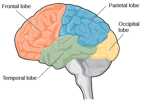
Figure 4. The lobes of the brain are shown.
People who suffer damage to Broca’s area have great difficulty producing language of any form. For example, Padma was an electrical engineer who was socially active and a caring, involved mother. About twenty years ago, she was in a car accident and suffered damage to her Broca’s area. She completely lost the ability to speak and form any kind of meaningful language. There is nothing wrong with her mouth or her vocal cords, but she is unable to produce words. She can follow directions but can’t respond verbally, and she can read but no longer write. She can do routine tasks like running to the market to buy milk, but she could not communicate verbally if a situation called for it.
One particularly fascinating area in the frontal lobe is called the “primary motor cortex.” This strip running along the side of the brain is in charge of voluntary movements like waving goodbye, wiggling your eyebrows, and kissing. It is an excellent example of the way that the various regions of the brain are highly specialized. Interestingly, each of our various body parts has a unique portion of the primary motor cortex devoted to it. Each individual finger has about as much dedicated brain space as your entire leg. Your lips, in turn, require about as much dedicated brain processing as all of your fingers and your hand combined!
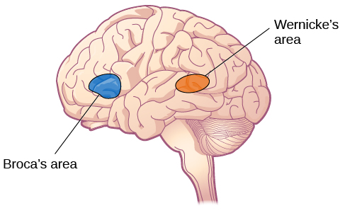
Figure 5. Damage to either Broca’s area or Wernicke’s area can result in language deficits. The types of deficits are very different, however, depending on which area is affected.
The temporal lobe is located on the side of the head (temporal means “near the temples”), and is associated with hearing, memory, emotion, and some aspects of language. The auditory cortex, the main area responsible for processing auditory information, is located within the temporal lobe. Wernicke’s area, important for speech comprehension, is also located here. Whereas individuals with damage to Broca’s area (frontal lobe) have difficulty producing language, those with damage to Wernicke’s area can produce sensible language, but they are unable to understand what others are saying.
The occipital lobe is located at the very back of the brain, and contains the primary visual cortex, which is responsible for interpreting incoming visual information. The occipital cortex is organized retinotopically, which means there is a close relationship between the position of an object in a person’s visual field and the position of that object’s representation on the cortex.
Try It
Watch It
So how does our understanding of the brain, the nervous system, and the endocrine apply to abnormal psychology? Biological psychologists seek to understand how genetic, biological, and physical components of the body and brain influence mental illness. Watch this video to see examples of the types of research that biological psychologists do, and specifically how research by Dr. Bonnie Nagel from Oregon Health & Science University is examining neurological structures and their connection to alcoholism and substance-abuse disorders.
You can view the transcript for “What is a Biological Psychologist?” here (opens in new window).
Glossary
auditory cortex: strip of cortex in the temporal lobe that is responsible for processing auditory information
autonomic nervous system: controls our internal organs and glands
Broca’s area: region in the left hemisphere frontal lobe that is essential for language production
central nervous system (CNS): brain and spinal cord
cerebral cortex: surface of the brain that is associated with our highest mental capabilities
fight-or-flight response: activation of the sympathetic division of the autonomic nervous system, allowing access to energy reserves and heightened sensory capacity so that we might fight off a given threat or run away to safety
forebrain: largest part of the brain; contains the cerebral cortex, the thalamus, and the limbic system, among other structures
frontal lobe: part of the cerebral cortex involved in reasoning, motor control, emotion, and language; contains motor cortex
hindbrain: lower end of the brainstem, mostly regulates autonomic body functions
homeostasis: state of equilibrium—biological conditions, such as body temperature, are maintained at optimal levels
hypothalamic-pituitary-adrenocortical (HPA) axis: pathway connecting the brain and the endocrine system, heavily involved in fight-or-flight reactions to threats
midbrain: top of the brainstem; coordinates sensory input and motor movement and regulates sleep/wake cycles
motor cortex: strip of cortex involved in planning and coordinating movement
occipital lobe: part of the cerebral cortex associated with visual processing; contains the primary visual cortex
parasympathetic nervous system: associated with routine, day-to-day operations of the body
parietal lobe: part of the cerebral cortex involved in processing various sensory and perceptual information; contains the primary somatosensory cortex
peripheral nervous system (PNS): connects the brain and spinal cord to the muscles, organs, and senses in the periphery of the body
prefrontal cortex: area in the frontal lobe responsible for higher-level cognitive functioning
somatic nervous system: relays sensory and motor information to and from the CNS
somatosensory cortex: essential for processing sensory information from across the body, such as touch, temperature, and pain
sulcus (plural: sulci): depressions or grooves in the cerebral cortex
sympathetic nervous system: involved in stress-related activities and functions
temporal lobe: part of cerebral cortex associated with hearing, memory, emotion, and some aspects of language; contains primary auditory cortex
Wernicke’s area: important for speech comprehension, located in the temporal lobe
Candela Citations
- Modification, adaptation, and original content. Authored by: Anton Tolman for Lumen Learning. Provided by: Lumen Learning. License: CC BY: Attribution
- The Brain and Spinal Cord. Provided by: OpenStax College. Located at: https://openstax.org/books/psychology-2e/pages/3-4-the-brain-and-spinal-cord. License: CC BY: Attribution. License Terms: Download for free at https://openstax.org/books/psychology-2e/pages/1-introduction.
- Motor cortex paragraphs and image. Authored by: Robert Biswas-Diener. Provided by: Portland State University. Located at: http://nobaproject.com/modules/the-brain-and-nervous-system. Project: The Noba Project. License: CC BY-NC-SA: Attribution-NonCommercial-ShareAlike
- Parts of the Nervous System. Provided by: OpenStax College. Located at: https://openstax.org/books/psychology-2e/pages/3-3-parts-of-the-nervous-system. License: CC BY: Attribution. License Terms: Download for free at https://openstax.org/books/psychology-2e/pages/1-introduction
- The Endocrine System. Provided by: OpenStax College. Located at: https://openstax.org/books/psychology-2e/pages/3-5-the-endocrine-system. License: CC BY: Attribution. License Terms: Download for free at https://openstax.org/books/psychology-2e/pages/1-introduction
- Sympathetic nervous system. Provided by: Wikipedia. Located at: https://en.wikipedia.org/wiki/Sympathetic_nervous_system. License: CC BY-SA: Attribution-ShareAlike
- What is a Biological Psychologist?. Provided by: Portland Community College. Located at: https://www.youtube.com/watch?time_continue=9&v=iEVtX6_99-U&feature=emb_logo. License: Other. License Terms: Standard YouTube License
- Durand, V.M. & Barlow, D.H. (2013). Essentials of Abnormal Psychology. Cengage Learning. ↵