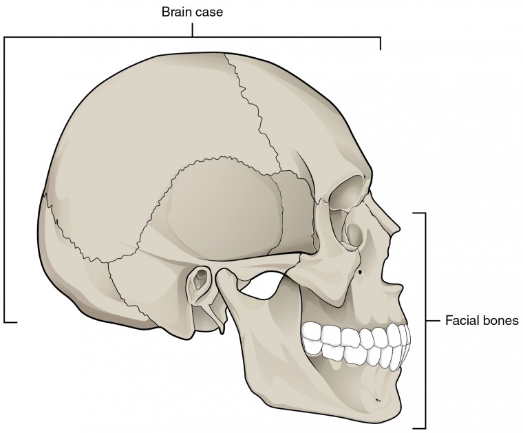
Figure 1: Parts of the Skull
The cranium (skull) is the skeletal structure of the head that supports the face and protects the brain. It is subdivided into the facial bones and the brain case. The facial bones underlie the facial structures, form the nasal cavity, enclose the eyeballs, and support the teeth of the upper and lower jaws. The rounded brain case surrounds and protects the brain and houses the middle and inner ear structures. In the adult, the skull consists of 22 individual bones, 21 of which are immobile and united into a single unit. The 22nd bone is the mandible (lower jaw), which is the only moveable bone of the skull.
Cranial Bones of the Skull:
Cranial bones form the cranial cavity that surrounds and protects the brain and brainstem. There are eight cranial bones. frontal, sphenoid, ethmoid, occipital, 2 temporal, and 2 parietal bones.These bones are important as they provide an articulation point for the 1st cervical vertebra (atlas), as well as the facial bones and the mandible (jaw bone). The bones that form the top and sides of the brain case are usually referred to as the “flat” bones of the skull.

Figure 2: Cranial Bones of Human Skull
- Frontal bone (1) – The frontal bone is the single bone that forms the forehead.
- Parietal bone (2) – The parietal bone forms most of the upper lateral side of the skull . These are paired bones, with the right and left parietal bones joining together at the top of the skull. They are examples of flat bones.
- Temporal bone (2) – The temporal bone forms the lower lateral side of the skull. Common wisdom has it that the temporal bone (temporal = “time”) is so named because this area of the head (the temple) is where hair typically first turns gray, indicating the passage of time. Two major structures if the temporal bone are the external auditory meatus and the mastoid process. The external auditory meatus (the ear canal) which is a prominent canal in the tympanic part of the temporal bone. It transmits sounds to the tympanic membrane (eardrum) in the middle ear. The mastoid process is a large prominence behind your earlobe which serves as a muscle attachment site
- Occipital bone (1) – The occipital bone is the single bone that forms the posterior skull and posterior base of the cranial cavity. On the base of the skull, the occipital bone contains the large opening of the foramen magnum, which allows for passage of the spinal cord as it exits the skull. On either side of the foramen magnum is an oval-shaped occipital condyle. These condyles form joints with the first cervical vertebra and thus support the skull on top of the vertebral column.
- Sphenoid bone (1) – The sphenoid bone is a single, complex bone of the central skull. It serves as a “keystone” bone, because it joins with almost every other bone of the skull. The sphenoid forms much of the base of the central skull and also extends laterally to contribute to the sides of the skull. Inside the cranial cavity, it has a pair of lesser wings and a pair of greater wings. The right and left lesser wings of the sphenoid bone, resemble the wings of a flying bird. The greater wings of the sphenoid bone extend laterally to either side. The greater wing is best seen on the outside of the lateral skull, where it forms a rectangular area immediately anterior to the temporal bone.
- Ethmoid bone: The ethmoid bone is a single, midline bone that forms the roof and lateral walls of the upper nasal cavity, the upper portion of the nasal septum, and contributes to the medial wall of the orbit . On the interior of the skull, the ethmoid also forms a portion of the floor of the anterior cranial cavity.
Sutures of the Skull

Figure 3: Immovable Suture Joints of Skull
A suture is an immobile joint between adjacent bones of the skull. The narrow gap between the bones is filled with dense, fibrous connective tissue that unites the bones. The long sutures located between the bones of the brain case are not straight, but instead follow irregular, tightly twisting paths. These twisting lines serve to tightly interlock the adjacent bones, thus adding strength to the skull for brain protection.
Fontanelles

Figure 4 (a) Superior view of infant skull.

Figure 4(b) Lateral view of infant skull:
A fontanelle is an anatomical feature on an infant’s skull that allows its plates to be flexible to pass through the birth canal. Fontanelles are soft spots on a baby’s head that, during birth, enable the bony plates of the skull to flex and allow the child’s head to pass through the birth canal. The ossification of the bones of the skull causes the fontanelles to close over a period of 18 to 24 months; they eventually form the sutures of the neurocranium. The cranium of a newborn consists of five main bones: two frontal bones, two parietal bones, and one occipital bone. These are joined by fibrous sutures that allow movement that facilitates childbirth and brain growth. At birth, the skull features a small posterior fontanelle (an open area covered by a tough membrane) where the two parietal bones adjoin the occipital bone (at the lambda). This fontanelle usually closes during the first two to three months of an infant’s life.
The much larger, diamond-shaped anterior fontanelle—where the two frontal and two parietal bones join—generally remains open until a child is about two years old. The anterior fontanelle is useful clinically, as examination of an infant includes palpating the anterior fontanelle. Two smaller fontanelles are located on each side of the head. The more anterior one is the sphenoidal (between the sphenoid, parietal, temporal, and frontal bones), while the more posterior one is the mastoid (between the temporal, occipital, and parietal bones).
The fontanelle may pulsate. Although the precise cause of this is not known, it is perfectly normal and seems to echo the heartbeat, perhaps via the arterial pulse within the brain vasculature, or in the meninges. This pulsating action is how the soft spot got its name: fontanelle means little fountain. Parents may worry that their infant may be more prone to injury at the fontanelles. In fact, although they may colloquially be called soft spots, the membrane covering the fontanelles is extremely tough and difficult to penetrate. The fontanelles allow the infant brain to be imaged using ultrasonography. Once they are closed, most of the brain is inaccessible to ultrasound imaging because the bony skull presents an acoustic barrier.
Facial Bones of the Skull
The facial bones of the skull form the upper and lower jaws, the nose, nasal cavity and nasal septum, and the orbit. The facial bones include 14 bones, with six paired bones and two unpaired bones. The paired bones are the maxilla, palatine, zygomatic, nasal, lacrimal, and inferior nasal conchae bones. The unpaired bones are the vomer and mandible bones. Although classified with the brain-case bones, the ethmoid bone also contributes to the nasal septum and the walls of the nasal cavity and orbit.

Figure 5: Facial Bones of Human Skull
- Zygomatic Bone (2) – Forms the cheek bones of the face, and articulates with the frontal, sphenoid, temporal and maxilla bones.
- Lacrimal Bone (2) – The smallest bones of the face. They form part of the medial wall of the orbit.
- Nasal Bone (2) – The nasal bone is one of two small bones that articulate (join) with each other to form the bony base (bridge) of the nose. They also support the cartilages that form the lateral walls of the nose. These are the bones that are damaged when the nose is broken.
- Inferior nasal conchae (2) – Located within the nasal cavity, these bones increase the surface area of the nasal cavity, thus increasing the amount of inspired air that can come into contact with the cavity walls.
- Palatine Bone (2) – Situated at the rear of oral cavity, and forms part of the hard palate.
- Maxilla (2) ( upper jaw bone) – Comprises part of the upper jaw and hard palate. The maxillary bone forms the upper jaw and supports the upper teeth. Each maxilla also forms the lateral floor of each orbit and the majority of the hard palate. The hard palate is the bony plate that forms the roof of the mouth and floor of the nasal cavity, separating the oral and nasal cavities.
- Vomer – The unpaired vomer bone, often referred to simply as the vomer, is triangular-shaped and forms the posterior-inferior part of the nasal septum. The vomer is best seen when looking from behind into the posterior openings of the nasal cavity . In this view, the vomer is seen to form the entire height of the nasal septum. A much smaller portion of the vomer can also be seen when looking into the anterior opening of the nasal cavity.
- Mandible ( lower jaw bone) – The mandible forms the lower jaw and is the only moveable bone of the skull. The posterior part of mandible is topped by the oval-shaped condyle. The condyle of the mandible articulates (joins) with the temporal bone. Together these articulations form the temporomandibular joint, which allows for opening and closing of the mouth. The broad U-shaped curve located between the coronoid and condylar processes is the mandibular notch.
HOMEOSTATIC IMBALANCES: CLEFT LIP AND CLEFT PALATE


During embryonic development, the right and left maxilla bones come together at the midline to form the upper jaw. At the same time, the muscle and skin overlying these bones join together to form the upper lip. Inside the mouth, the palatine processes of the maxilla bones, along with the horizontal plates of the right and left palatine bones, join together to form the hard palate. If an error occurs in these developmental processes, a birth defect of cleft lip or cleft palate may result.
Cleft lip is a common development defect that affects approximately 1:1000 births, most of which are male. This defect involves a partial or complete failure of the right and left portions of the upper lip to fuse together, leaving a cleft (gap).
A more severe developmental defect is cleft palate, which affects the hard palate. The hard palate is the bony structure that separates the nasal cavity from the oral cavity. It is formed during embryonic development by the midline fusion of the horizontal plates from the right and left palatine bones and the palatine processes of the maxilla bones. Cleft palate affects approximately 1:2500 births and is more common in females. It results from a failure of the two halves of the hard palate to completely come together and fuse at the midline, thus leaving a gap between them. This gap allows for communication between the nasal and oral cavities. In severe cases, the bony gap continues into the anterior upper jaw where the alveolar processes of the maxilla bones also do not properly join together above the front teeth. If this occurs, a cleft lip will also be seen. Because of the communication between the oral and nasal cavities, a cleft palate makes it very difficult for an infant to generate the suckling needed for nursing, thus leaving the infant at risk for malnutrition. Surgical repair is required to correct cleft palate defects.
Paranasal Sinuses
The paranasal sinuses are hollow, air-filled spaces located within certain bones of the skull. All of the sinuses communicate with the nasal cavity (paranasal = “next to nasal cavity”) and are lined with nasal mucosa. Their function is to reduce bone mass and thus lighten the skull, and they also add resonance to the voice. This second feature is most obvious when you have a cold or sinus congestion. These produce swelling of the mucosa and excess mucus production, which can obstruct the narrow passageways between the sinuses and the nasal cavity, causing your voice to sound different to yourself and others. This blockage can also allow the sinuses to fill with fluid, with the resulting pressure producing pain and discomfort.

Figure 6: Paranasal Sinuses
The paranasal sinuses are named for the skull bone that each occupies. The frontal sinus is located just above the eyebrows, within the frontal bone. This irregular space may be divided at the midline into bilateral spaces, or these may be fused into a single sinus space. The frontal sinus is the most anterior of the paranasal sinuses. The largest sinus is the maxillary sinus. These are paired and located within the right and left maxillary bones, where they occupy the area just below the orbits. The maxillary sinuses are most commonly involved during sinus infections. Because their connection to the nasal cavity is located high on their medial wall, they are difficult to drain. The sphenoid sinus is a single, midline sinus. It is located within the body of the sphenoid bone, just anterior and inferior to the sella turcica, thus making it the most posterior of the paranasal sinuses. The lateral aspects of the ethmoid bone contain multiple small spaces separated by very thin bony walls. Each of these spaces is called an ethmoid air cell. These are located on both sides of the ethmoid bone, between the upper nasal cavity and medial orbit, just behind the superior nasal conchae.
Hyoid Bone
The hyoid bone is an independent bone that does not contact any other bone and thus is not part of the skull. It is a small U-shaped bone located in the upper neck near the level of the inferior mandible, with the tips of the “U” pointing posteriorly. The hyoid serves as the base for the tongue above, and is attached to the larynx below and the pharynx posteriorly. The hyoid is held in position by a series of small muscles that attach to it either from above or below. These muscles act to move the hyoid up/down or forward/back. Movements of the hyoid are coordinated with movements of the tongue, larynx, and pharynx during swallowing and speaking.

Figure 7: Hyoid Bone.