The function of the digestive system is to break down the foods you eat, release their nutrients, and absorb those nutrients into the body. Although the small intestine is where the majority of digestion occurs, and where most of the released nutrients are absorbed into the blood or lymph, each of the digestive system organs makes a vital contribution to this process.
The processes of digestion include six activities:

Figure 1. Peristalsis moves food through the digestive tract with alternating waves of muscle contraction and relaxation.
- 1. Ingestion: The first of these processes, ingestion, refers to the entry of food into the alimentary canal through the mouth. Chewing increases the surface area of the food and allows an appropriately sized bolus to be produced.
- 2. Propulsion: Food leaves the mouth when the tongue and pharyngeal muscles propel it into the esophagus. This act of swallowing, the last voluntary act until defecation, is an example of propulsion, which refers to the movement of food through the digestive tract. It includes both the voluntary process of swallowing and the involuntary process of peristalsis. Peristalsis consists of sequential, alternating waves of contraction and relaxation of alimentary wall smooth muscles, which act to propel food along (Figure 1). These waves also play a role in mixing food with digestive juices. Peristalsis is so powerful that foods and liquids you swallow enter your stomach even if you are standing on your head.
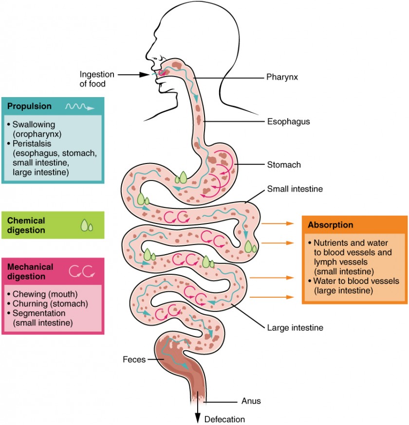
Figure 2. :The different processes involved in digestion.
- 3. Mechanical or physical digestion: Digestion includes both mechanical and chemical processes. Mechanical digestion is a purely physical process that does not change the chemical nature of the food. Instead, it makes the food smaller to increase both surface area and mobility. It includes mastication or chewing, mechanical churning of food in the stomach and segmentation in small intestine.
- 4. Chemical digestion : Chemical digestion, starts in the mouth, digestive secretions break down complex food molecules into their chemical building blocks (for example, proteins into separate amino acids). These secretions vary in composition, but typically contain water, various enzymes, acids, and salts. Chemical digestion is completed in the small intestine.
- 5. Absorption: Food that has been broken down is of no value to the body unless it enters the bloodstream and its nutrients are put to work. This occurs through the process of absorption, which takes place primarily within the small intestine.
- 6. Defecation / Elimination: In defecation, the final step in digestion, undigested materials are removed from the body as feces.
Digestive System Anatomy:

Figure 3. Mains organs of digestive system.
The human digestive system consists of a digestive tube, the alimentary canal, and accessory organs that secrete digestive chemicals.
Alimentary Canal and Organs: Also called the gastrointestinal (GI) tract or gut, the alimentary canal (aliment- = “to nourish”) is a one-way tube about 7.62 meters (25 feet) in length. The main function of the organs of the alimentary canal is to nourish the body. This tube begins at the mouth and terminates at the anus. Between those two points,the alimentary canal is divided into specialized digestive organs along its length :
mouth (oral cavity)→ pharynx→esophagus→ stomach→ small intestine→ large intestine (colon)→ rectum→ anus.
The inside space of the alimentary canal (tube) is called lumen. Food goes through the lumen. Both the mouth and anus are open to the external environment; thus, food and wastes within the alimentary canal are technically considered to be outside the body. Only through the process of absorption do the nutrients in food enter into and nourish the body’s “inner space.”
Wall of the Alimentary Canal
Throughout its length, the alimentary tract is composed of the same four tissue layers; the details of their structural arrangements vary to fit their specific functions. Starting from the lumen and moving outwards, these layers are the mucosa, submucosa, muscularis, and serosa.

Figure 4. The wall of the alimentary canal has four basic tissue layers: the mucosa, submucosa, muscularis, and serosa.
The mucosa is referred to as a mucous membrane, because mucus production is a characteristic feature of gut epithelium. The membrane consists of epithelium, which is in direct contact with ingested food.
The submucosa lies immediately beneath the mucosa. A broad layer of dense connective tissue, it connects the overlying mucosa to the underlying muscularis. It includes blood and lymphatic vessels (which transport absorbed nutrients), and a scattering of submucosal glands that release digestive secretions. Additionally, it serves as a conduit for a dense branching network of nerves.
The muscalaris (also called the muscularis externa) is the third layer of the alimentary canal. The muscularis in the small intestine is made up of a double layer of smooth muscle: an inner circular layer and an outer longitudinal layer. The contractions of these layers promote mechanical digestion, and move the food along the canal. The basic two-layer structure found in the small intestine is modified in the organs proximal and distal to it. The stomach is equipped for its churning function by the addition of a third layer, the oblique muscle. While the colon has two layers like the small intestine, its longitudinal layer is segregated into three narrow parallel bands, the tenia coli, which make it look like a series of pouches rather than a simple tube.
The serosa is the portion of the alimentary canal superficial to the muscularis. Present only in the region of the alimentary canal within the abdominal cavity, it consists of a layer of visceral peritoneum overlying a layer of loose connective tissue. Instead of serosa, the mouth, pharynx, and esophagus have a dense sheath of collagen fibers called the adventitia. These tissues serve to hold the alimentary canal in place near the ventral surface of the vertebral column.
Candela Citations
- Anatomy & Physiology. Provided by: OpenStax CNX. Located at: http://cnx.org/contents/14fb4ad7-39a1-4eee-ab6e-3ef2482e3e22@15.1.. License: CC BY: Attribution. License Terms: Download for free at http://cnx.org/contents/14fb4ad7-39a1-4eee-ab6e-3ef2482e3e22@8.25
- Mar 23, 2016 OpenStax. Textbook content produced by OpenStax is licensed under a Creative Commons Attribution License 4.0 license.. Provided by: OpenStax CNX. Located at: http://cnx.org/contents/185cbf87-c72e-48f5-b51e-f14f21b5eabd@11.6.. Project: Biology. License: CC BY: Attribution. License Terms: Creative Commons Attribution License 4.0 license.