What you’ll learn to do: identify the basic structures of a neuron, the function of each structure, and how messages travel through the neuron

Neuron in tissue culture.
Ever wonder how your brain actually works? What exactly is going on inside of your small, wrinkly mass while you read this text? In this section, you’ll learn about the basics of neural communication in the brain, which is the brain’s way of sending messages to and from different regions in order to relay critical information about your body and its surroundings.
Glia and neurons are the two cell types that make up the nervous system. While glia generally play supporting roles, the communication between neurons is fundamental to all of the functions associated with the nervous system. Neuronal communication is made possible by the neuron’s specialized structures, like the soma, dendrites, axons, terminal buttons, and synaptic vesicles.
Neuronal communication is an electrochemical event. The dendrites contain receptors for neurotransmitters released by nearby neurons. If the signals received from other neurons are sufficiently strong, an action potential will travel down the length of the axon to the terminal buttons, resulting in the release of neurotransmitters into the synapse.
Different neurotransmitters are associated with different functions. Often, psychological disorders involve imbalances in a given neurotransmitter system. Therefore, psychotropic drugs are prescribed in an attempt to bring the neurotransmitters back into balance. Drugs can act either as agonists or as antagonists for a given neurotransmitter system.
Learning Objectives
- Explain the role and function of the basic structures of a neuron
- Describe how neurons communicate with each other
- Explain how drugs act as agonists or antagonists for a given neurotransmitter system
Neurons
Psychologists striving to understand the human mind may study the nervous system. Learning how the cells and organs (like the brain) function, help us understand the biological basis behind human psychology. The nervous system is composed of two basic cell types: glial cells (also known as glia) and neurons. Glial cells, which outnumber neurons ten to one, are traditionally thought to play a supportive role to neurons, both physically and metabolically. Glial cells provide scaffolding on which the nervous system is built, help neurons line up closely with each other to allow neuronal communication, provide insulation to neurons, transport nutrients and waste products, and mediate immune responses. Neurons, on the other hand, serve as interconnected information processors that are essential for all of the tasks of the nervous system. This section briefly describes the structure and function of neurons.
Neuron Structure
Neurons are the central building blocks of the nervous system, 100 billion strong at birth. Like all cells, neurons consist of several different parts, each serving a specialized function (Figure 1). A neuron’s outer surface is made up of a semipermeable membrane. This membrane allows smaller molecules and molecules without an electrical charge to pass through it, while stopping larger or highly charged molecules.
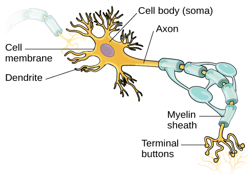
Figure 1. This illustration shows a prototypical neuron, which is being myelinated.
The nucleus of the neuron is located in the soma, or cell body. The soma has branching extensions known as dendrites. The neuron is a small information processor, and dendrites serve as input sites where signals are received from other neurons. These signals are transmitted electrically across the soma and down a major extension from the soma known as the axon, which ends at multiple terminal buttons. The terminal buttons contain synaptic vesicles that house neurotransmitters, the chemical messengers of the nervous system.
Axons range in length from a fraction of an inch to several feet. In some axons, glial cells form a fatty substance known as the myelin sheath, which coats the axon and acts as an insulator, increasing the speed at which the signal travels. The myelin sheath is crucial for the normal operation of the neurons within the nervous system: the loss of the insulation it provides can be detrimental to normal function. To understand how this works, let’s consider an example. Multiple sclerosis (MS), an autoimmune disorder, involves a large-scale loss of the myelin sheath on axons throughout the nervous system. The resulting interference in the electrical signal prevents the quick transmittal of information by neurons and can lead to a number of symptoms, such as dizziness, fatigue, loss of motor control, and sexual dysfunction. While some treatments may help to modify the course of the disease and manage certain symptoms, there is currently no known cure for multiple sclerosis.
In healthy individuals, the neuronal signal moves rapidly down the axon to the terminal buttons, where synaptic vesicles release neurotransmitters into the synapse (Figure 2). The synapse is a very small space between two neurons and is an important site where communication between neurons occurs. Once neurotransmitters are released into the synapse, they travel across the small space and bind with corresponding receptors on the dendrite of an adjacent neuron. Receptors, proteins on the cell surface where neurotransmitters attach, vary in shape, with different shapes “matching” different neurotransmitters.
How does a neurotransmitter “know” which receptor to bind to? The neurotransmitter and the receptor have what is referred to as a lock-and-key relationship—specific neurotransmitters fit specific receptors similar to how a key fits a lock. The neurotransmitter binds to any receptor that it fits.
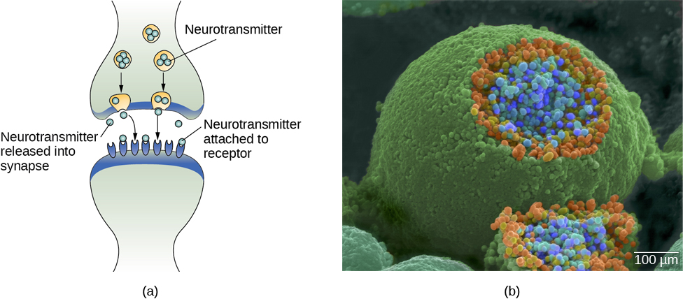
Figure 2. (a) The synapse is the space between the terminal button of one neuron and the dendrite of another neuron. (b) In this pseudo-colored image from a scanning electron microscope, a terminal button (green) has been opened to reveal the synaptic vesicles (orange and blue) inside. Each vesicle contains about 10,000 neurotransmitter molecules. (credit b: modification of work by Tina Carvalho, NIH-NIGMS; scale-bar data from Matt Russell)
Link to Learning
Click through the links at the top of this interactive simulation to review the parts of a nerve cell and to take a closer look at how neurons communicate.
Try It
Glossary
Candela Citations
- Modification, adaptation, and original content. Provided by: Lumen Learning. License: CC BY: Attribution
- Addition of link to learning. Provided by: Lumen Learning. License: CC BY: Attribution
- Neuron in tissue culture. Located at: https://commons.wikimedia.org/wiki/File:Neuron_in_tissue_culture.jpg. License: CC BY-SA: Attribution-ShareAlike
- Cells of the Nervous System. Authored by: OpenStax College. Located at: http://cnx.org/contents/Sr8Ev5Og@5.52:SO2ufnKm@7/Cells-of-the-Nervous-System. License: CC BY: Attribution. License Terms: Download for free at http://cnx.org/contents/4abf04bf-93a0-45c3-9cbc-2cefd46e68cc@5.48
- 2-Minute Neuroscience: The Neuron. Authored by: Neuroscientifically Challenged. Located at: https://www.youtube.com/watch?v=6qS83wD29PY. License: Other. License Terms: Standard YouTube License