Chapter 14. DNA Structure and Function

Introduction*
The three letters “DNA” have now become synonymous with crime solving, paternity testing, human identification, and genetic testing. DNA can be retrieved from hair, blood, or saliva. Each person’s DNA is unique, and it is possible to detect differences between individuals within a species on the basis of these unique features.
DNA analysis has many practical applications beyond forensics. In humans, DNA testing is applied to numerous uses: determining paternity, tracing genealogy, identifying pathogens, archeological research, tracing disease outbreaks, and studying human migration patterns. In the medical field, DNA is used in diagnostics, new vaccine development, and cancer therapy. It is now possible to determine predisposition to diseases by looking at genes.
Each human cell has 23 pairs of chromosomes: one set of chromosomes is inherited from the mother and the other set is inherited from the father. There is also a mitochondrial genome, inherited exclusively from the mother, which can be involved in inherited genetic disorders. On each chromosome, there are thousands of genes that are responsible for determining the genotype and phenotype of the individual. A gene is defined as a sequence of DNA that codes for a functional product. The human haploid genome contains 3 billion base pairs and has between 20,000 and 25,000 functional genes.
14.1. Historical Basis of Modern Understanding*
By the end of this section, you will be able to:
-
Explain transformation of DNA
-
Describe the key experiments that helped identify that DNA is the genetic material
-
State and explain Chargaff’s rules
Modern understandings of DNA have evolved from the discovery of nucleic acid to the development of the double-helix model. In the 1860s, Friedrich Miescher (Figure 14.2), a physician by profession, was the first person to isolate phosphate-rich chemicals from white blood cells or leukocytes. He named these chemicals (which would eventually be known as RNA and DNA) nuclein because they were isolated from the nuclei of the cells.


To see Miescher conduct an experiment step-by-step, click through this review of how he discovered the key role of DNA and proteins in the nucleus.
A half century later, British bacteriologist Frederick Griffith was perhaps the first person to show that hereditary information could be transferred from one cell to another “horizontally,” rather than by descent. In 1928, he reported the first demonstration of bacterial transformation, a process in which external DNA is taken up by a cell, thereby changing morphology and physiology. He was working with Streptococcus pneumoniae, the bacterium that causes pneumonia. Griffith worked with two strains, rough (R) and smooth (S). The R strain is non-pathogenic (does not cause disease) and is called rough because its outer surface is a cell wall and lacks a capsule; as a result, the cell surface appears uneven under the microscope. The S strain is pathogenic (disease-causing) and has a capsule outside its cell wall. As a result, it has a smooth appearance under the microscope. Griffith injected the live R strain into mice and they survived. In another experiment, when he injected mice with the heat-killed S strain, they also survived. In a third set of experiments, a mixture of live R strain and heat-killed S strain were injected into mice, and—to his surprise—the mice died. Upon isolating the live bacteria from the dead mouse, only the S strain of bacteria was recovered. When this isolated S strain was injected into fresh mice, the mice died. Griffith concluded that something had passed from the heat-killed S strain into the live R strain and transformed it into the pathogenic S strain, and he called this the transforming principle (Figure 14.3). These experiments are now famously known as Griffith’s transformation experiments.

Scientists Oswald Avery, Colin MacLeod, and Maclyn McCarty (1944) were interested in exploring this transforming principle further. They isolated the S strain from the dead mice and isolated the proteins and nucleic acids, namely RNA and DNA, as these were possible candidates for the molecule of heredity. They conducted a systematic elimination study. They used enzymes that specifically degraded each component and then used each mixture separately to transform the R strain. They found that when DNA was degraded, the resulting mixture was no longer able to transform the bacteria, whereas all of the other combinations were able to transform the bacteria. This led them to conclude that DNA was the transforming principle.
DNA evidence was used for the first time to solve an immigration case. The story started with a teenage boy returning to London from Ghana to be with his mother. Immigration authorities at the airport were suspicious of him, thinking that he was traveling on a forged passport. After much persuasion, he was allowed to go live with his mother, but the immigration authorities did not drop the case against him. All types of evidence, including photographs, were provided to the authorities, but deportation proceedings were started nevertheless. Around the same time, Dr. Alec Jeffreys of Leicester University in the United Kingdom had invented a technique known as DNA fingerprinting. The immigration authorities approached Dr. Jeffreys for help. He took DNA samples from the mother and three of her children, plus an unrelated mother, and compared the samples with the boy’s DNA. Because the biological father was not in the picture, DNA from the three children was compared with the boy’s DNA. He found a match in the boy’s DNA for both the mother and his three siblings. He concluded that the boy was indeed the mother’s son.
Forensic scientists analyze many items, including documents, handwriting, firearms, and biological samples. They analyze the DNA content of hair, semen, saliva, and blood, and compare it with a database of DNA profiles of known criminals. Analysis includes DNA isolation, sequencing, and sequence analysis; most forensic DNA analysis involves polymerase chain reaction (PCR) amplification of short tandem repeat (STR) loci and electrophoresis to determine the length of the PCR-amplified fragment. Only mitochondrial DNA is sequenced for forensics. Forensic scientists are expected to appear at court hearings to present their findings. They are usually employed in crime labs of city and state government agencies. Geneticists experimenting with DNA techniques also work for scientific and research organizations, pharmaceutical industries, and college and university labs. Students wishing to pursue a career as a forensic scientist should have at least a bachelor’s degree in chemistry, biology, or physics, and preferably some experience working in a laboratory.
Experiments conducted by Martha Chase and Alfred Hershey in 1952 provided confirmatory evidence that DNA was the genetic material and not proteins. Chase and Hershey were studying a bacteriophage, which is a virus that infects bacteria. Viruses typically have a simple structure: a protein coat, called the capsid, and a nucleic acid core that contains the genetic material, either DNA or RNA. The bacteriophage infects the host bacterial cell by attaching to its surface, and then it injects its nucleic acids inside the cell. The phage DNA makes multiple copies of itself using the host machinery, and eventually the host cell bursts, releasing a large number of bacteriophages. Hershey and Chase labeled one batch of phage with radioactive sulfur, 35S, to label the protein coat. Another batch of phage were labeled with radioactive phosphorus, 32P. Because phosphorous is found in DNA, but not protein, the DNA and not the protein would be tagged with radioactive phosphorus.
Each batch of phage was allowed to infect the cells separately. After infection, the phage bacterial suspension was put in a blender, which caused the phage coat to be detached from the host cell. The phage and bacterial suspension was spun down in a centrifuge. The heavier bacterial cells settled down and formed a pellet, whereas the lighter phage particles stayed in the supernatant. In the tube that contained phage labeled with 35S, the supernatant contained the radioactively labeled phage, whereas no radioactivity was detected in the pellet. In the tube that contained the phage labeled with 32P, the radioactivity was detected in the pellet that contained the heavier bacterial cells, and no radioactivity was detected in the supernatant. Hershey and Chase concluded that it was the phage DNA that was injected
into the cell and carried information to produce more phage particles, thus providing evidence that DNA was the genetic material and not proteins (Figure 14.4).

Around this same time, Austrian biochemist Erwin Chargaff examined the content of DNA in different species and found that the amounts of adenine, thymine, guanine, and cytosine were not found in equal quantities, and that it varied from species to species, but not between individuals of the same species. He found that the amount of adenine equals the amount of thymine, and the amount of cytosine equals the amount of guanine, or A = T and G = C. This is also known as Chargaff’s rules. This finding proved immensely useful when Watson and Crick were getting ready to propose their DNA double helix model.
14.2. DNA Structure and Sequencing*
By the end of this section, you will be able to:
-
Describe the structure of DNA
-
Explain the Sanger method of DNA sequencing
-
Discuss the similarities and differences between eukaryotic and prokaryotic DNA
The building blocks of DNA are nucleotides. The important components of the nucleotide are a nitrogenous base, deoxyribose (5-carbon sugar), and a phosphate group (Figure 14.5). The nucleotide is named depending on the nitrogenous base. The nitrogenous base can be a purine such as adenine (A) and guanine (G), or a pyrimidine such as cytosine (C) and thymine (T).

The nucleotides combine with each other by covalent bonds known as phosphodiester bonds or linkages. The purines have a double ring structure with a six-membered ring fused to a five-membered ring. Pyrimidines are smaller in size; they have a single six-membered ring structure. The carbon atoms of the five-carbon sugar are numbered 1′, 2′, 3′, 4′, and 5′ (1′ is read as “one prime”). The phosphate residue is attached to the hydroxyl group of the 5′ carbon of one sugar of one nucleotide and the hydroxyl group of the 3′ carbon of the sugar of the next nucleotide, thereby forming a 5′-3′ phosphodiester bond.
In the 1950s, Francis Crick and James Watson worked together to determine the structure of DNA at the University of Cambridge, England. Other scientists like Linus Pauling and Maurice Wilkins were also actively exploring this field. Pauling had discovered the secondary structure of proteins using X-ray crystallography. In Wilkins’ lab, researcher Rosalind Franklin was using X-ray diffraction methods to understand the structure of DNA. Watson and Crick were able to piece together the puzzle of the DNA molecule on the basis of Franklin’s data because Crick had also studied X-ray diffraction (Figure 14.6). In 1962, James Watson, Francis Crick, and Maurice Wilkins were awarded the Nobel Prize in Medicine. Unfortunately, by then Franklin had died, and Nobel prizes are not awarded posthumously.

Watson and Crick proposed that DNA is made up of two strands that are twisted around each other to form a right-handed helix. Base pairing takes place between a purine and pyrimidine; namely, A pairs with T and G pairs with C. Adenine and thymine are complementary base pairs, and cytosine and guanine are also complementary base pairs. The base pairs are stabilized by hydrogen bonds; adenine and thymine form two hydrogen bonds and cytosine and guanine form three hydrogen bonds. The two strands are anti-parallel in nature; that is, the 3′ end of one strand faces the 5′ end of the other strand. The sugar and phosphate of the nucleotides form the backbone of the structure, whereas the nitrogenous bases are stacked inside. Each base pair is separated from the other base pair by a distance of 0.34 nm, and each turn of the helix measures 3.4 nm. Therefore, ten base pairs are present per turn of the helix. The diameter of the DNA double helix is 2 nm, and it is uniform throughout. Only the pairing between a purine and pyrimidine can explain the uniform diameter. The twisting of the two strands around each other results in the formation of uniformly spaced major and minor grooves (Figure 14.7).

DNA Sequencing Techniques
Until the 1990s, the sequencing of DNA (reading the sequence of DNA) was a relatively expensive and long process. Using radiolabeled nucleotides also compounded the problem through safety concerns. With currently available technology and automated machines, the process is cheap, safer, and can be completed in a matter of hours. Fred Sanger developed the sequencing method used for the human genome sequencing project, which is widely used today (Figure 14.8).

Visit this site to watch a video explaining the DNA sequence reading technique that resulted from Sanger’s work.
The method is known as the dideoxy chain termination method. The sequencing method is based on the use of chain terminators, the dideoxynucleotides (ddNTPs). The dideoxynucleotides, or ddNTPSs, differ from the deoxynucleotides by the lack of a free 3′ OH group on the five-carbon sugar. If a ddNTP is added to a growing a DNA strand, the chain is not extended any further because the free 3′ OH group needed to add another nucleotide is not available. By using a predetermined ratio of deoxyribonucleotides to dideoxynucleotides, it is possible to generate DNA fragments of different sizes.

The DNA sample to be sequenced is denatured or separated into two strands by heating it to high temperatures. The DNA is divided into four tubes in which a primer, DNA polymerase, and all four nucleotides (A, T, G, and C) are added. In addition to each of the four tubes, limited quantities of one of the four dideoxynucleotides are added to each tube respectively. The tubes are labeled as A, T, G, and C according to the ddNTP added. For detection purposes, each of the four dideoxynucleotides carries a different fluorescent label. Chain elongation continues until a fluorescent dideoxy nucleotide is incorporated, after which no further elongation takes place. After the reaction is over, electrophoresis is performed. Even a difference in length of a single base can be detected. The sequence is read from a laser scanner. For his work on DNA sequencing, Sanger received a Nobel Prize in chemistry in 1980.

Sanger’s genome sequencing has led to a race to sequence human genomes at a rapid speed and low cost, often referred to as the $1000 in one day sequence. Learn more by selecting the Sequencing at Speed animation here.
Gel electrophoresis is a technique used to separate DNA fragments of different sizes. Usually the gel is made of a chemical called agarose. Agarose powder is added to a buffer and heated. After cooling, the gel solution is poured into a casting tray. Once the gel has solidified, the DNA is loaded on the gel and electric current is applied. The DNA has a net negative charge and moves from the negative electrode toward the positive electrode. The electric current is applied for sufficient time to let the DNA separate according to size; the smallest fragments will be farthest from the well (where the DNA was loaded), and the heavier molecular weight fragments will be closest to the well. Once the DNA is separated, the gel is stained with a DNA-specific dye for viewing it (Figure 14.9).

The first draft sequence of the Neanderthal genome was recently published by Richard E. Green et al. in 2010.[11] Neanderthals are the closest ancestors of present-day humans. They were known to have lived in Europe and Western Asia before they disappeared from fossil records approximately 30,000 years ago. Green’s team studied almost 40,000-year-old fossil remains that were selected from sites across the world. Extremely sophisticated means of sample preparation and DNA sequencing were employed because of the fragile nature of the bones and heavy microbial contamination. In their study, the scientists were able to sequence some four billion base pairs. The Neanderthal sequence was compared with that of present-day humans from across the world. After comparing the sequences, the researchers found that the Neanderthal genome had 2 to 3 percent greater similarity to people living outside Africa than to people in Africa. While current theories have suggested that all present-day humans can be traced to a small ancestral population in Africa, the data from the Neanderthal genome may contradict this view. Green and his colleagues also discovered DNA segments among people in Europe and Asia that are more similar to Neanderthal sequences than to other contemporary human sequences. Another interesting observation was that Neanderthals are as closely related to people from Papua New Guinea as to those from China or France. This is surprising because Neanderthal fossil remains have been located only in Europe and West Asia. Most likely, genetic exchange took place between Neanderthals and modern humans as modern humans emerged out of Africa, before the divergence of Europeans, East Asians, and Papua New Guineans.
Several genes seem to have undergone changes from Neanderthals during the evolution of present-day humans. These genes are involved in cranial structure, metabolism, skin morphology, and cognitive development. One of the genes that is of particular interest is RUNX2, which is different in modern day humans and Neanderthals. This gene is responsible for the prominent frontal bone, bell-shaped rib cage, and dental differences seen in Neanderthals. It is speculated that an evolutionary change in RUNX2 was important in the origin of modern-day humans, and this affected the cranium and the upper body.

Watch Svante Pääbo’s talk explaining the Neanderthal genome research at the 2011 annual TED (Technology, Entertainment, Design) conference.
DNA Packaging in Cells
When comparing prokaryotic cells to eukaryotic cells, prokaryotes are much simpler than eukaryotes in many of their features (Figure 14.10). Most prokaryotes contain a single, circular chromosome that is found in an area of the cytoplasm called the nucleoid.

In eukaryotic cells, DNA and RNA synthesis occur in a separate compartment from protein synthesis. In prokaryotic cells, both processes occur together. What advantages might there be to separating the processes? What advantages might there be to having them occur together?
The size of the genome in one of the most well-studied prokaryotes, E.coli, is 4.6 million base pairs (approximately 1.1 mm, if cut and stretched out). So how does this fit inside a small bacterial cell? The DNA is twisted by what is known as supercoiling. Supercoiling means that DNA is either under-wound (less than one turn of the helix per 10 base pairs) or over-wound (more than 1 turn per 10 base pairs) from its normal relaxed state. Some proteins are known to be involved in the supercoiling; other proteins and enzymes such as DNA gyrase help in maintaining the supercoiled structure.
Eukaryotes, whose chromosomes each consist of a linear DNA molecule, employ a different type of packing strategy to fit their DNA inside the nucleus (Figure 14.11). At the most basic level, DNA is wrapped around proteins known as histones to form structures called nucleosomes. The histones are evolutionarily conserved proteins that are rich in basic amino acids and form an octamer. The DNA (which is negatively charged because of the phosphate groups) is wrapped tightly around the histone core. This nucleosome is linked to the next one with the help of a linker DNA. This is also known as the “beads on a string” structure. This is further compacted into a 30 nm fiber, which is the diameter of the structure. At the metaphase stage, the chromosomes are at their most compact, are approximately 700 nm in width, and are found in association with scaffold proteins.
In interphase, eukaryotic chromosomes have two distinct regions that can be distinguished by staining. The tightly packaged region is known as heterochromatin, and the less dense region is known as euchromatin. Heterochromatin usually contains genes that are not expressed, and is found in the regions of the centromere and telomeres. The euchromatin usually contains genes that are transcribed, with DNA packaged around nucleosomes but not further compacted.

14.3. Basics of DNA Replication*
By the end of this section, you will be able to:
-
Explain how the structure of DNA reveals the replication process
-
Describe the Meselson and Stahl experiments
The elucidation of the structure of the double helix provided a hint as to how DNA divides and makes copies of itself. This model suggests that the two strands of the double helix separate during replication, and each strand serves as a template from which the new complementary strand is copied. What was not clear was how the replication took place. There were three models suggested (Figure 14.12): conservative, semi-conservative, and dispersive.

In conservative replication, the parental DNA remains together, and the newly formed daughter strands are together. The semi-conservative method suggests that each of the two parental DNA strands act as a template for new DNA to be synthesized; after replication, each double-stranded DNA includes one parental or “old” strand and one “new” strand. In the dispersive model, both copies of DNA have double-stranded segments of parental DNA and newly synthesized DNA interspersed.
Meselson and Stahl were interested in understanding how DNA replicates. They grew E. coli for several generations in a medium containing a “heavy” isotope of nitrogen (15N) that gets incorporated into nitrogenous bases, and eventually into the DNA (Figure 14.13).

The E. coli culture was then shifted into medium containing 14N and allowed to grow for one generation. The cells were harvested and the DNA was isolated. The DNA was centrifuged at high speeds in an ultracentrifuge. Some cells were allowed to grow for one more life cycle in 14N and spun again. During the density gradient centrifugation, the DNA is loaded into a gradient (typically a salt such as cesium chloride or sucrose) and spun at high speeds of 50,000 to 60,000 rpm. Under these circumstances, the DNA will form a band according to its density in the gradient. DNA grown in 15N will band at a higher density position than that grown in 14N. Meselson and Stahl noted that after one generation of growth in 14N after they had been shifted from 15N, the single band observed was intermediate in position in between DNA of cells grown exclusively in 15N and 14N. This suggested either a semi-conservative or dispersive mode of replication. The DNA harvested from cells grown for two generations in 14N formed two bands: one DNA band was at the intermediate position between 15N and 14N, and the other corresponded to the band of 14N DNA. These results could only be explained if DNA replicates in a semi-conservative manner. Therefore, the other two modes were ruled out.
During DNA replication, each of the two strands that make up the double helix serves as a template from which new strands are copied. The new strand will be complementary to the parental or “old” strand. When two daughter DNA copies are formed, they have the same sequence and are divided equally into the two daughter cells.

Click through this tutorial on DNA replication.
14.4. DNA Replication in Prokaryotes*
By the end of this section, you will be able to:
-
Explain the process of DNA replication in prokaryotes
-
Discuss the role of different enzymes and proteins in supporting this process
DNA replication has been extremely well studied in prokaryotes primarily because of the small size of the genome and the mutants that are available. E. coli has 4.6 million base pairs in a single circular chromosome and all of it gets replicated in approximately 42 minutes, starting from a single origin of replication and proceeding around the circle in both directions. This means that approximately 1000 nucleotides are added per second. The process is quite rapid and occurs without many mistakes.
DNA replication employs a large number of proteins and enzymes, each of which plays a critical role during the process. One of the key players is the enzyme DNA polymerase, also known as DNA pol, which adds nucleotides one by one to the growing DNA chain that are complementary to the template strand. The addition of nucleotides requires energy; this energy is obtained from the nucleotides that have three phosphates attached to them, similar to ATP which has three phosphate groups attached. When the bond between the phosphates is broken, the energy released is used to form the phosphodiester bond between the incoming nucleotide and the growing chain. In prokaryotes, three main types of polymerases are known: DNA pol I, DNA pol II, and DNA pol III. It is now known that DNA pol III is the enzyme required for DNA synthesis; DNA pol I and DNA pol II are primarily required for repair.
How does the replication machinery know where to begin? It turns out that there are specific nucleotide sequences called origins of replication where replication begins. In E. coli, which has a single origin of replication on its one chromosome (as do most prokaryotes), it is approximately 245 base pairs long and is rich in AT sequences. The origin of replication is recognized by certain proteins that bind to this site. An enzyme called helicase unwinds the DNA by breaking the hydrogen bonds between the nitrogenous base pairs. ATP hydrolysis is required for this process. As the DNA opens up, Y-shaped structures called replication forks are formed. Two replication forks are formed at the origin of replication and these get extended bi- directionally as replication proceeds. Single-strand binding proteins coat the single strands of DNA near the replication fork to prevent the single-stranded DNA from winding back into a double helix. DNA polymerase is able to add nucleotides only in the 5′ to 3′ direction (a new DNA strand can be only extended in this direction). It also requires a free 3′-OH group to which it can add nucleotides by forming a phosphodiester bond between the 3′-OH end and the 5′ phosphate of the next nucleotide. This essentially means that it cannot add nucleotides if a free 3′-OH group is not available. Then how does it add the first nucleotide? The problem is solved with the help of a primer that provides the free 3′-OH end. Another enzyme, RNA primase, synthesizes an RNA primer that is about five to ten nucleotides long and complementary to the DNA. Because this sequence primes the DNA synthesis, it is appropriately called the primer. DNA polymerase can now extend this RNA primer, adding nucleotides one by one that are complementary to the template strand (Figure 14.14).

You isolate a cell strain in which the joining together of Okazaki fragments is impaired and suspect that a mutation has occurred in an enzyme found at the replication fork. Which enzyme is most likely to be mutated?
The replication fork moves at the rate of 1000 nucleotides per second. DNA polymerase can only extend in the 5′ to 3′ direction, which poses a slight problem at the replication fork. As we know, the DNA double helix is anti-parallel; that is, one strand is in the 5′ to 3′ direction and the other is oriented in the 3′ to 5′ direction. One strand, which is complementary to the 3′ to 5′ parental DNA strand, is synthesized continuously towards the replication fork because the polymerase can add nucleotides in this direction. This continuously synthesized strand is known as the leading strand. The other strand, complementary to the 5′ to 3′ parental DNA, is extended away from the replication fork, in small fragments known as Okazaki fragments, each requiring a primer to start the synthesis. Okazaki fragments are named after the Japanese scientist who first discovered them. The strand with the Okazaki fragments is known as the lagging strand.
The leading strand can be extended by one primer alone, whereas the lagging strand needs a new primer for each of the short Okazaki fragments. The overall direction of the lagging strand will be 3′ to 5′, and that of the leading strand 5′ to 3′. A protein called the sliding clamp holds the DNA polymerase in place as it continues to add nucleotides. The sliding clamp is a ring-shaped protein that binds to the DNA and holds the polymerase in place. Topoisomerase prevents the over-winding of the DNA double helix ahead of the replication fork as the DNA is opening up; it does so by causing temporary nicks in the DNA helix and then resealing it. As synthesis proceeds, the RNA primers are replaced by DNA. The primers are removed by the exonuclease activity of DNA pol I, and the gaps are filled in by deoxyribonucleotides. The nicks that remain between the newly synthesized DNA (that replaced the RNA primer) and the previously synthesized DNA are sealed by the enzyme DNA ligase that catalyzes the formation of phosphodiester linkage between the 3′-OH end of one nucleotide and the 5′ phosphate end of the other fragment.
Once the chromosome has been completely replicated, the two DNA copies move into two different cells during cell division. The process of DNA replication can be summarized as follows:
-
DNA unwinds at the origin of replication.
-
Helicase opens up the DNA-forming replication forks; these are extended bidirectionally.
-
Single-strand binding proteins coat the DNA around the replication fork to prevent rewinding of the DNA.
-
Topoisomerase binds at the region ahead of the replication fork to prevent supercoiling.
-
Primase synthesizes RNA primers complementary to the DNA strand.
-
DNA polymerase starts adding nucleotides to the 3′-OH end of the primer.
-
Elongation of both the lagging and the leading strand continues.
-
RNA primers are removed by exonuclease activity.
-
Gaps are filled by DNA pol by adding dNTPs.
-
The gap between the two DNA fragments is sealed by DNA ligase, which helps in the formation of phosphodiester bonds.
<!–cnx.newline–>
Table 14.1 summarizes the enzymes involved in prokaryotic DNA replication and the functions of each.
| Prokaryotic DNA Replication: Enzymes and Their Function | |
|---|---|
| Enzyme/protein | Specific Function |
| DNA pol I | Exonuclease activity removes RNA primer and replaces with newly synthesized DNA |
| DNA pol II | Repair function |
| DNA pol III | Main enzyme that adds nucleotides in the 5′-3′ direction |
| Helicase | Opens the DNA helix by breaking hydrogen bonds between the nitrogenous bases |
| Ligase | Seals the gaps between the Okazaki fragments to create one continuous DNA strand |
| Primase | Synthesizes RNA primers needed to start replication |
| Sliding Clamp | Helps to hold the DNA polymerase in place when nucleotides are being added |
| Topoisomerase | Helps relieve the stress on DNA when unwinding by causing breaks and then resealing the DNA |
| Single-strand binding proteins (SSB) | Binds to single-stranded DNA to avoid DNA rewinding back. |

Review the full process of DNA replication here.
14.5. DNA Replication in Eukaryotes*
By the end of this section, you will be able to:
-
Discuss the similarities and differences between DNA replication in eukaryotes and prokaryotes
-
State the role of telomerase in DNA replication
Eukaryotic genomes are much more complex and larger in size than prokaryotic genomes. The human genome has three billion base pairs per haploid set of chromosomes, and 6 billion base pairs are replicated during the S phase of the cell cycle. There are multiple origins of replication on the eukaryotic chromosome; humans can have up to 100,000 origins of replication. The rate of replication is approximately 100 nucleotides per second, much slower than prokaryotic replication. In yeast, which is a eukaryote, special sequences known as Autonomously Replicating Sequences (ARS) are found on the chromosomes. These are equivalent to the origin of replication in E. coli.
The number of DNA polymerases in eukaryotes is much more than prokaryotes: 14 are known, of which five are known to have major roles during replication and have been well studied. They are known as pol α, pol β, pol γ, pol δ, and pol ε.
The essential steps of replication are the same as in prokaryotes. Before replication can start, the DNA has to be made available as template. Eukaryotic DNA is bound to basic proteins known as histones to form structures called nucleosomes. The chromatin (the complex between DNA and proteins) may undergo some chemical modifications, so that the DNA may be able to slide off the proteins or be accessible to the enzymes of the DNA replication machinery. At the origin of replication, a pre-replication complex is made with other initiator proteins. Other proteins are then recruited to start the replication process (Table 14.2).
A helicase using the energy from ATP hydrolysis opens up the DNA helix. Replication forks are formed at each replication origin as the DNA unwinds. The opening of the double helix causes over-winding, or supercoiling, in the DNA ahead of the replication fork. These are resolved with the action of topoisomerases. Primers are formed by the enzyme primase, and using the primer, DNA pol can start synthesis. While the leading strand is continuously synthesized by the enzyme pol δ, the lagging strand is synthesized by pol ε. A sliding clamp protein known as PCNA (Proliferating Cell Nuclear Antigen) holds the DNA pol in place so that it does not slide off the DNA. RNase H removes the RNA primer, which is then replaced with DNA nucleotides. The Okazaki fragments in the lagging strand are joined together after the replacement of the RNA primers with DNA. The gaps that remain are sealed by DNA ligase, which forms the phosphodiester bond.
Telomere replication
Unlike prokaryotic chromosomes, eukaryotic chromosomes are linear. As you’ve learned, the enzyme DNA pol can add nucleotides only in the 5′ to 3′ direction. In the leading strand, synthesis continues until the end of the chromosome is reached. On the lagging strand, DNA is synthesized in short stretches, each of which is initiated by a separate primer. When the replication fork reaches the end of the linear chromosome, there is no place for a primer to be made for the DNA fragment to be copied at the end of the chromosome. These ends thus remain unpaired, and over time these ends may get progressively shorter as cells continue to divide.
The ends of the linear chromosomes are known as telomeres, which have repetitive sequences that code for no particular gene. In a way, these telomeres protect the genes from getting deleted as cells continue to divide. In humans, a six base pair sequence, TTAGGG, is repeated 100 to 1000 times. The discovery of the enzyme telomerase (Figure 14.16) helped in the understanding of how chromosome ends are maintained. The telomerase enzyme contains a catalytic part and a built-in RNA template. It attaches to the end of the chromosome, and complementary bases to the RNA template are added on the 3′ end of the DNA strand. Once the 3′ end of the lagging strand template is sufficiently elongated, DNA polymerase can add the nucleotides complementary to the ends of the chromosomes. Thus, the ends of the chromosomes are replicated.

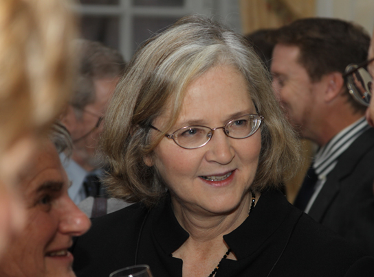
Telomerase is typically active in germ cells and adult stem cells. It is not active in adult somatic cells. For her discovery of telomerase and its action, Elizabeth Blackburn (Figure 14.16) received the Nobel Prize for Medicine and Physiology in 2009.
Telomerase and Aging
Cells that undergo cell division continue to have their telomeres shortened because most somatic cells do not make telomerase. This essentially means that telomere shortening is associated with aging. With the advent of modern medicine, preventative health care, and healthier lifestyles, the human life span has increased, and there is an increasing demand for people to look younger and have a better quality of life as they grow older.
In 2010, scientists found that telomerase can reverse some age-related conditions in mice. This may have potential in regenerative medicine.[12] Telomerase-deficient mice were used in these studies; these mice have tissue atrophy, stem cell depletion, organ system failure, and impaired tissue injury responses. Telomerase reactivation in these mice caused extension of telomeres, reduced DNA damage, reversed neurodegeneration, and improved the function of the testes, spleen, and intestines. Thus, telomere reactivation may have potential for treating age-related diseases in humans.
Cancer is characterized by uncontrolled cell division of abnormal cells. The cells accumulate mutations, proliferate uncontrollably, and can migrate to different parts of the body through a process called metastasis. Scientists have observed that cancerous cells have considerably shortened telomeres and that telomerase is active in these cells. Interestingly, only after the telomeres were shortened in the cancer cells did the telomerase become active. If the action of telomerase in these cells can be inhibited by drugs during cancer therapy, then the cancerous cells could potentially be stopped from further division.
| Difference between Prokaryotic and Eukaryotic Replication | ||
|---|---|---|
| Property | Prokaryotes | Eukaryotes |
| Origin of replication | Single | Multiple |
| Rate of replication | 1000 nucleotides/s | 50 to 100 nucleotides/s |
| DNA polymerase types | 5 | 14 |
| Telomerase | Not present | Present |
| RNA primer removal | DNA pol I | RNase H |
| Strand elongation | DNA pol III | Pol δ, pol ε |
| Sliding clamp | Sliding clamp | PCNA |
14.6. DNA Repair*
By the end of this section, you will be able to:
-
Discuss the different types of mutations in DNA
-
Explain DNA repair mechanisms
DNA replication is a highly accurate process, but mistakes can occasionally occur, such as a DNA polymerase inserting a wrong base. Uncorrected mistakes may sometimes lead to serious consequences, such as cancer. Repair mechanisms correct the mistakes. In rare cases, mistakes are not corrected, leading to mutations; in other cases, repair enzymes are themselves mutated or defective.
Most of the mistakes during DNA replication are promptly corrected by DNA polymerase by proofreading the base that has been just added (Figure 14.17). In proofreading, the DNA pol reads the newly added base before adding the next one, so a correction can be made. The polymerase checks whether the newly added base has paired correctly with the base in the template strand. If it is the right base, the next nucleotide is added. If an incorrect base has been added, the enzyme makes a cut at the phosphodiester bond and releases the wrong nucleotide. This is performed by the exonuclease action of DNA pol III. Once the incorrect nucleotide has been removed, a new one will be added again.

Some errors are not corrected during replication, but are instead corrected after replication is completed; this type of repair is known as mismatch repair (Figure 14.18). The enzymes recognize the incorrectly added nucleotide and excise it; this is then replaced by the correct base. If this remains uncorrected, it may lead to more permanent damage. How do mismatch repair enzymes recognize which of the two bases is the incorrect one? In E. coli, after replication, the nitrogenous base adenine acquires a methyl group; the parental DNA strand will have methyl groups, whereas the newly synthesized strand lacks them. Thus, DNA polymerase is able to remove the wrongly incorporated bases from the newly synthesized, non-methylated strand. In eukaryotes, the mechanism is not very well understood, but it is believed to involve recognition of unsealed nicks in the new strand, as well as a short-term continuing association of some of the replication proteins with the new daughter strand after replication has completed.

In another type of repair mechanism, nucleotide excision repair, enzymes replace incorrect bases by making a cut on both the 3′ and 5′ ends of the incorrect base (Figure 14.19). The segment of DNA is removed and replaced with the correctly paired nucleotides by the action of DNA pol. Once the bases are filled in, the remaining gap is sealed with a phosphodiester linkage catalyzed by DNA ligase. This repair mechanism is often employed when UV exposure causes the formation of pyrimidine dimers.

A well-studied example of mistakes not being corrected is seen in people suffering from xeroderma pigmentosa (Figure 14.20). Affected individuals have skin that is highly sensitive to UV rays from the sun. When individuals are exposed to UV, pyrimidine dimersespecially those of thymineare formed; people with xeroderma pigmentosa are not able to repair the damage. These are not repaired because of a defect in the nucleotide excision repair enzymes, whereas in normal individuals, the thymine dimers are excised and the defect is corrected. The thymine dimers distort the structure of the DNA double helix, and this may cause problems during DNA replication. People with xeroderma pigmentosa may have a higher risk of contracting skin cancer than those who dont have the condition.

Errors during DNA replication are not the only reason why mutations arise in DNA. Mutations, variations in the nucleotide sequence of a genome, can also occur because of damage to DNA. Such mutations may be of two types: induced or spontaneous. Induced mutations are those that result from an exposure to chemicals, UV rays, x-rays, or some other environmental agent. Spontaneous mutations occur without any exposure to any environmental agent; they are a result of natural reactions taking place within the body.
Mutations may have a wide range of effects. Some mutations are not expressed; these are known as silent mutations. Point mutations are those mutations that affect a single base pair. The most common nucleotide mutations are substitutions, in which one base is replaced by another. These can be of two types, either transitions or transversions. Transition substitution refers to a purine or pyrimidine being replaced by a base of the same kind; for example, a purine such as adenine may be replaced by the purine guanine. Transversion substitution refers to a purine being replaced by a pyrimidine, or vice versa; for example, cytosine, a pyrimidine, is replaced by adenine, a purine. Mutations can also be the result of the addition of a base, known as an insertion, or the removal of a base, also known as deletion. Sometimes a piece of DNA from one chromosome may get translocated to another chromosome or to another region of the same chromosome; this is also known as translocation. These mutation types are shown in Figure 14.21.

A frameshift mutation that results in the insertion of three nucleotides is often less deleterious than a mutation that results in the insertion of one nucleotide. Why?
Mutations in repair genes have been known to cause cancer. Many mutated repair genes have been implicated in certain forms of pancreatic cancer, colon cancer, and colorectal cancer. Mutations can affect either somatic cells or germ cells. If many mutations accumulate in a somatic cell, they may lead to problems such as the uncontrolled cell division observed in cancer. If a mutation takes place in germ cells, the mutation will be passed on to the next generation, as in the case of hemophilia and xeroderma pigmentosa.
Glossary
- electrophoresis
-
technique used to separate DNA fragments according to size
- helicase
-
during replication, this enzyme helps to open up the DNA helix by breaking the hydrogen bonds
- induced mutation
-
mutation that results from exposure to chemicals or environmental agents
- lagging strand
-
during replication, the strand that is replicated in short fragments and away from the replication fork
- leading strand
-
strand that is synthesized continuously in the 5′-3′ direction which is synthesized in the direction of the replication fork
- ligase
-
enzyme that catalyzes the formation of a phosphodiester linkage between the 3′ OH and 5′ phosphate ends of the DNA
- mismatch repair
-
type of repair mechanism in which mismatched bases are removed after replication
- mutation
-
variation in the nucleotide sequence of a genome
- nucleotide excision repair
-
type of DNA repair mechanism in which the wrong base, along with a few nucleotides upstream or downstream, are removed
- Okazaki fragment
-
DNA fragment that is synthesized in short stretches on the lagging strand
- point mutation
-
mutation that affects a single base
- primase
-
enzyme that synthesizes the RNA primer; the primer is needed for DNA pol to start synthesis of a new DNA strand
- primer
-
short stretch of nucleotides that is required to initiate replication; in the case of replication, the primer has RNA nucleotides
- proofreading
-
function of DNA pol in which it reads the newly added base before adding the next one
- replication fork
-
Y-shaped structure formed during initiation of replication
- silent mutation
-
mutation that is not expressed
- single-strand binding protein
-
during replication, protein that binds to the single-stranded DNA; this helps in keeping the two strands of DNA apart so that they may serve as templates
- sliding clamp
-
ring-shaped protein that holds the DNA pol on the DNA strand
- spontaneous mutation
-
mutation that takes place in the cells as a result of chemical reactions taking place naturally without exposure to any external agent
- telomerase
-
enzyme that contains a catalytic part and an inbuilt RNA template; it functions to maintain telomeres at chromosome ends
- telomere
-
DNA at the end of linear chromosomes
- topoisomerase
-
enzyme that causes underwinding or overwinding of DNA when DNA replication is taking place
- transformation
-
process in which external DNA is taken up by a cell
- transition substitution
-
when a purine is replaced with a purine or a pyrimidine is replaced with another pyrimidine
- transversion substitution
-
when a purine is replaced by a pyrimidine or a pyrimidine is replaced by a purine
<!–CNX: Start Area: “Sections Summary”–>
DNA was first isolated from white blood cells by Friedrich Miescher, who called it nuclein because it was isolated from nuclei. Frederick Griffith’s experiments with strains of Streptococcus pneumoniae provided the first hint that DNA may be the transforming principle. Avery, MacLeod, and McCarty proved that DNA is required for the transformation of bacteria. Later experiments by Hershey and Chase using bacteriophage T2 proved that DNA is the genetic material. Chargaff found that the ratio of A = T and C = G, and that the percentage content of A, T, G, and C is different for different species.
The currently accepted model of the double-helix structure of DNA was proposed by Watson and Crick. Some of the salient features are that the two strands that make up the double helix are complementary and anti-parallel in nature. Deoxyribose sugars and phosphates form the backbone of the structure, and the nitrogenous bases are stacked inside. The diameter of the double helix, 2 nm, is uniform throughout. A purine always pairs with a pyrimidine; A pairs with T, and G pairs with C. One turn of the helix has ten base pairs. During cell division, each daughter cell receives a copy of the DNA by a process known as DNA replication. Prokaryotes are much simpler than eukaryotes in many of their features. Most prokaryotes contain a single, circular chromosome. In general, eukaryotic chromosomes contain a linear DNA molecule packaged into nucleosomes, and have two distinct regions that can be distinguished by staining, reflecting different states of packaging and compaction.
The model for DNA replication suggests that the two strands of the double helix separate during replication, and each strand serves as a template from which the new complementary strand is copied. In conservative replication, the parental DNA is conserved, and the daughter DNA is newly synthesized. The semi-conservative method suggests that each of the two parental DNA strands acts as template for new DNA to be synthesized; after replication, each double-stranded DNA includes one parental or “old” strand and one “new” strand. The dispersive mode suggested that the two copies of the DNA would have segments of parental DNA and newly synthesized DNA.
Replication in prokaryotes starts from a sequence found on the chromosome called the origin of replication—the point at which the DNA opens up. Helicase opens up the DNA double helix, resulting in the formation of the replication fork. Single-strand binding proteins bind to the single-stranded DNA near the replication fork to keep the fork open. Primase synthesizes an RNA primer to initiate synthesis by DNA polymerase, which can add nucleotides only in the 5′ to 3′ direction. One strand is synthesized continuously in the direction of the replication fork; this is called the leading strand. The other strand is synthesized in a direction away from the replication fork, in short stretches of DNA known as Okazaki fragments. This strand is known as the lagging strand. Once replication is completed, the RNA primers are replaced by DNA nucleotides and the DNA is sealed with DNA ligase, which creates phosphodiester bonds between the 3′-OH of one end and the 5′ phosphate of the other strand.
Replication in eukaryotes starts at multiple origins of replication. The mechanism is quite similar to prokaryotes. A primer is required to initiate synthesis, which is then extended by DNA polymerase as it adds nucleotides one by one to the growing chain. The leading strand is synthesized continuously, whereas the lagging strand is synthesized in short stretches called Okazaki fragments. The RNA primers are replaced with DNA nucleotides; the DNA remains one continuous strand by linking the DNA fragments with DNA ligase. The ends of the chromosomes pose a problem as polymerase is unable to extend them without a primer. Telomerase, an enzyme with an inbuilt RNA template, extends the ends by copying the RNA template and extending one end of the chromosome. DNA polymerase can then extend the DNA using the primer. In this way, the ends of the chromosomes are protected.
DNA polymerase can make mistakes while adding nucleotides. It edits the DNA by proofreading every newly added base. Incorrect bases are removed and replaced by the correct base, and then a new base is added. Most mistakes are corrected during replication, although when this does not happen, the mismatch repair mechanism is employed. Mismatch repair enzymes recognize the wrongly incorporated base and excise it from the DNA, replacing it with the correct base. In yet another type of repair, nucleotide excision repair, the incorrect base is removed along with a few bases on the 5′ and 3′ end, and these are replaced by copying the template with the help of DNA polymerase. The ends of the newly synthesized fragment are attached to the rest of the DNA using DNA ligase, which creates a phosphodiester bond.
Most mistakes are corrected, and if they are not, they may result in a mutation defined as a permanent change in the DNA sequence. Mutations can be of many types, such as substitution, deletion, insertion, and translocation. Mutations in repair genes may lead to serious consequences such as cancer. Mutations can be induced or may occur spontaneously.
<!–CNX: Start Area: “Art Connections”–>
Figure 14.10 In eukaryotic cells, DNA and RNA synthesis occur in a separate compartment from protein synthesis. In prokaryotic cells, both processes occur together. What advantages might there be to separating the processes? What advantages might there be to having them occur together?
Figure 14.14 You isolate a cell strain in which the joining together of Okazaki fragments is impaired and suspect that a mutation has occurred in an enzyme found at the replication fork. Which enzyme is most likely to be mutated?
Figure 14.14 DNA ligase, as this enzyme joins together Okazaki fragments.
Figure 14.21 A frameshift mutation that results in the insertion of three nucleotides is often less deleterious than a mutation that results in the insertion of one nucleotide. Why?
<!–CNX: Start Area: “Multiple Choice”–>
If DNA of a particular species was analyzed and it was found that it contains 27 percent A, what would be the percentage of C?
-
27 percent
-
30 percent
-
23 percent
-
54 percent
C
The experiments by Hershey and Chase helped confirm that DNA was the hereditary material on the basis of the finding that:
-
radioactive phage were found in the pellet
-
radioactive cells were found in the supernatant
-
radioactive sulfur was found inside the cell
-
radioactive phosphorus was found in the cell
D
DNA double helix does not have which of the following?
-
antiparallel configuration
-
complementary base pairing
-
major and minor grooves
-
uracil
D
In eukaryotes, what is the DNA wrapped around?
-
single-stranded binding proteins
-
sliding clamp
-
polymerase
-
histones
D
Meselson and Stahl’s experiments proved that DNA replicates by which mode?
-
conservative
-
semi-conservative
-
dispersive
-
none of the above
B
If the sequence of the 5′-3′ strand is AATGCTAC, then the complementary sequence has the following sequence:
-
3′-AATGCTAC-5′
-
3′-CATCGTAA-5′
-
3′-TTACGATG-5′
-
3′-GTAGCATT-5′
C
Which of the following components is not involved during the formation of the replication fork?
-
single-strand binding proteins
-
helicase
-
origin of replication
-
ligase
D
Which of the following does the enzyme primase synthesize?
-
DNA primer
-
RNA primer
-
Okazaki fragments
-
phosphodiester linkage
B
In which direction does DNA replication take place?
-
5′-3′
-
3′-5′
-
5′
-
3′
A
The ends of the linear chromosomes are maintained by
-
helicase
-
primase
-
DNA pol
-
telomerase
D
During proofreading, which of the following enzymes reads the DNA?
-
primase
-
topoisomerase
-
DNA pol
-
helicase
C
The initial mechanism for repairing nucleotide errors in DNA is ________.
-
mismatch repair
-
DNA polymerase proofreading
-
nucleotide excision repair
-
thymine dimers
B
<!–CNX: Start Area: “Free Response”–>
Explain Griffith’s transformation experiments. What did he conclude from them?
Live R cells acquired genetic information from the heat-killed S cells that “transformed” the R cells into S cells.
Why were radioactive sulfur and phosphorous used to label bacteriophage in Hershey and Chase’s experiments?
Sulfur is an element found in proteins and phosphorus is a component of nucleic acids.
Provide a brief summary of the Sanger sequencing method.
The template DNA strand is mixed with a DNA polymerase, a primer, the 4 deoxynucleotides, and a limiting concentration of 4 dideoxynucleotides. DNA polymerase synthesizes a strand complementary to the template. Incorporation of ddNTPs at different locations results in DNA fragments that have terminated at every possible base in the template. These fragments are separated by gel electrophoresis and visualized by a laser detector to determine the sequence of bases.
Describe the structure and complementary base pairing of DNA.
DNA has two strands in anti-parallel orientation. The sugar-phosphate linkages form a backbone on the outside, and the bases are paired on the inside: A with T, and G with C, like rungs on a spiral ladder.
How did the scientific community learn that DNA replication takes place in a semi-conservative fashion?
Meselson’s experiments with E. coli grown in 15N deduced this finding.
DNA replication is bidirectional and discontinuous; explain your understanding of those concepts.
At an origin of replication, two replication forks are formed that are extended in two directions. On the lagging strand, Okazaki fragments are formed in a discontinuous manner.
What are Okazaki fragments and how they are formed?
Short DNA fragments are formed on the lagging strand synthesized in a direction away from the replication fork. These are synthesized by DNA pol.
If the rate of replication in a particular prokaryote is 900 nucleotides per second, how long would it take 1.2 million base pair genomes to make two copies?
1333 seconds or 22.2 minutes.
Explain the events taking place at the replication fork. If the gene for helicase is mutated, what part of replication will be affected?
At the replication fork, the events taking place are helicase action, binding of single-strand binding proteins, primer synthesis, and synthesis of new strands. If there is a mutated helicase gene, the replication fork will not be extended.
What is the role of a primer in DNA replication? What would happen if you forgot to add a primer in a tube containing the reaction mix for a DNA sequencing reaction?
Primer provides a 3′-OH group for DNA pol to start adding nucleotides. There would be no reaction in the tube without a primer, and no bands would be visible on the electrophoresis.
How do the linear chromosomes in eukaryotes ensure that its ends are replicated completely?
Telomerase has an inbuilt RNA template that extends the 3′ end, so primer is synthesized and extended. Thus, the ends are protected.
What is the consequence of mutation of a mismatch repair enzyme? How will this affect the function of a gene?
Mutations are not repaired, as in the case of xeroderma pigmentosa. Gene function may be affected or it may not be expressed.
<!–CNX: Start Area: “Art Connections”–>
Figure 14.14 DNA ligase, as this enzyme joins together Okazaki fragments.
<!–CNX: Start Area: “Multiple Choice”–>
C
D
D
D
B
C
D
B
A
D
C
B
<!–CNX: Start Area: “Free Response”–>
Live R cells acquired genetic information from the heat-killed S cells that “transformed” the R cells into S cells.
Sulfur is an element found in proteins and phosphorus is a component of nucleic acids.
The template DNA strand is mixed with a DNA polymerase, a primer, the 4 deoxynucleotides, and a limiting concentration of 4 dideoxynucleotides. DNA polymerase synthesizes a strand complementary to the template. Incorporation of ddNTPs at different locations results in DNA fragments that have terminated at every possible base in the template. These fragments are separated by gel electrophoresis and visualized by a laser detector to determine the sequence of bases.
DNA has two strands in anti-parallel orientation. The sugar-phosphate linkages form a backbone on the outside, and the bases are paired on the inside: A with T, and G with C, like rungs on a spiral ladder.
Meselson’s experiments with E. coli grown in 15N deduced this finding.
At an origin of replication, two replication forks are formed that are extended in two directions. On the lagging strand, Okazaki fragments are formed in a discontinuous manner.
Short DNA fragments are formed on the lagging strand synthesized in a direction away from the replication fork. These are synthesized by DNA pol.
1333 seconds or 22.2 minutes.
At the replication fork, the events taking place are helicase action, binding of single-strand binding proteins, primer synthesis, and synthesis of new strands. If there is a mutated helicase gene, the replication fork will not be extended.
Primer provides a 3′-OH group for DNA pol to start adding nucleotides. There would be no reaction in the tube without a primer, and no bands would be visible on the electrophoresis.
Telomerase has an inbuilt RNA template that extends the 3′ end, so primer is synthesized and extended. Thus, the ends are protected.
Mutations are not repaired, as in the case of xeroderma pigmentosa. Gene function may be affected or it may not be expressed.