Learning Outcomes
- Identify the structure and function of the reproductive system
In simple terms, reproduction is the process by which organisms create descendants. This miracle is a characteristic that all living things have in common and sets them apart from nonliving things. But even though the reproductive system is essential to keeping a species alive, it is not essential to keeping an individual alive.
In human reproduction, two kinds of sex cells or gametes are involved. Sperm, the male gamete, and a secondary oocyte (along with first polar body and corona radiata), the female gamete must meet in the female reproductive system to create a new individual. For reproduction to occur, both the female and male reproductive systems are essential. It is a common misnomer to refer to a woman’s gametic cell as an egg or ovum, but this is impossible. A secondary oocyte must be fertilized by the male gamete before it becomes an “ovum” or “egg.”
While both the female and male reproductive systems are involved with producing, nourishing and transporting either the oocyte or sperm, they are different in shape and structure. The male has reproductive organs, or genitals, that are both inside and outside the pelvis, while the female has reproductive organs entirely within the pelvis.
The Male Reproductive System
The male reproductive system consists of the testes and a series of ducts and glands. Sperm are produced in the testes and are transported through the reproductive ducts. These ducts include the epididymis, vas deferens, ejaculatory duct and urethra. The reproductive glands produce secretions that become part of semen, the fluid that is ejaculated from the urethra. These glands include the seminal vesicles, prostate gland, and bulbourethral glands.

Figure 1. The reproductive structures of the human male are shown.
Table 1 describes the major components of the male reproductive system.
| Table 1. Components of the Male Reproductive System | ||
|---|---|---|
| Structure | Location & Description | Function |
| Bulbourethral glands (2) | Pea sized organs posterior to the prostate on either side of the urethra. | Secretion of gelatinous seminal fluid called pre-ejaculate. This fluid helps to lubricate the urethra for spermatozoa to pass through, and to help flush out any residual urine or foreign matter. (< 1% of semen) |
| Epididymis | Tightly coiled duct lying just outside each testis connecting efferent ducts to vas deferens. | Storage and maturation of sperm. |
| Penis | Three columns of erectile tissue: two corpora cavernosa and one corpus spongiosum. Urethra passes through penis. | Male reproductive organ and also male organ of urination. |
| Prostate gland | Surrounds the urethra just below the urinary bladder and can be felt during a rectal exam. | Stores and secretes a clear, slightly alkaline fluid constituting up to one-third of the volume of semen. Raise vaginal pH.(25-30% of semen) |
| Seminal vesicles (2) | Convoluted structure attached to vas deferens near the base of the urinary bladder. | About 65-75% of the seminal fluid in humans originates from the seminal vesicles. Contain proteins, enzymes, fructose, mucus, vitamin C, flavins, phosphorylcholine and prostaglandins. High fructose concentrations provide nutrient energy for the spermatozoa as they travel through the female reproductive system. |
| Testes | Inside scrotum, outside of body. | Gonads that produce sperm and male sex hormones.Production of testosterone by cells of Leydig in the testicles. |
| Urethra | Connects bladder to outside body, about 8 inches long. | Tubular structure that receives urine from bladder and carries it to outside of the body. Also passage for sperm. |
| Vas deferens | Muscular tubes connecting the left and right epididymis to the ejaculatory ducts to move sperm. Each tube is about 30 cm long. | During ejaculation the smooth muscle in the vas deferens wall contracts, propelling sperm forward. Sperm are transferred from the vas deferens into the urethra, collecting fluids from accessory sex glands en route |
The Female Reproductive System
Reproduction can be defined as the process by which an organism continues its species. As noted earlier, in the human reproductive process, two kinds of gametes are involved: the male gamete (sperm) and the female gamete (egg or ovum). These two gametes meet within the female’s uterine tubes located one on each side of the upper pelvic cavity, and begin to create a new individual. The female needs a male to fertilize her egg; she then carries offspring through pregnancy and childbirth.
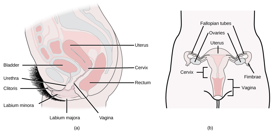
Figure 2. The reproductive structures of the human female are shown. (credit a: modification of work by Gray’s Anatomy; credit b: modification of work by CDC)
Female Reproductive System
- Produces eggs (ova)
- Secretes sex hormones
- Receives the male spermatozoa during
- Protects and nourishes the fertilized egg until it is fully developed
- Delivers fetus through birth canal
- Provides nourishment to the baby through milk secreted by mammary glands in the breast
The major components of the female reproductive system are shown in Table 2.
| Table 2. Components of the Male Reproductive System | ||
|---|---|---|
| Structure | Location & Description | Function |
| Ovaries (2) | Ovoid structures on either side of the uterus in the pelvic cavity | Primary sex organs of female; contain ovarian follicles that contain the oocytes. Oocytes are released during the ovulation stage of the menstrual cycle. |
| Fallopian Tubes (2) | Extend from lateral areas of the uterus to near the ovaries | Transport oocyte to uterus after fertilization and are the sites where fertilization by sperm actually occurs |
| Uterus | Pear shaped structure divided into the fundus and the cervix | Site of fetal development during gestation |
| Vagina | Located between rectum and urethra; smooth muscle lined with an epithelial mucous membrane | Path for menstrual blood and tissue to leave the body, as well as the fetus during childbirth. Produces a variety of secretions for lubrication and receives secretions that facilitate fertilization. |
| Vulva | Externally located: labia majora and minora, mons pubis, clithoris, vestibule, greater and lesser vestibular glands | Sexual function: heavily innervated and provide pleasure when properly stimulated. |
| Perineum | Area between vagina and anus | Helps form the muscular floor of pelvis; can be torn during vaginal childbirth |
| Mammary glands | Superficial to pectoral muscles | Provide nourishment to the baby through milk secretions |
Comparing Male and Female Reproductive Systems
Similarities
The reproductive systems of the male and female have some basic similarities and some specialized differences. They are the same in that most of the reproductive organs of both sexes develop from similar embryonic tissue, meaning they are homologous. Both systems have gonads that produce (sperm and egg or ovum) and sex organs. And both systems experience maturation of their reproductive organs, which become functional during puberty as a result of the gonads secreting sex hormones.
| Table 3. | ||
|---|---|---|
| Indifferent | Male | Female |
| Gonad | Testis | Ovary |
| Müllerian duct | Appendix testis | Fallopian tubes |
| Müllerian duct | Prostatic utricle | Uterus, proximal vagina |
| Wolffian duct | Rete testis | Rete ovarii |
| Mesonephric tubules | Efferent ducts | Epoophoron |
| Wolffian duct | Epididymis | Gartner’s duct |
| Wolffian duct | Vas deferens | |
| Wolffian duct | Seminal vesicle | |
| Wolffian duct | Prostate | Skene’s glands |
| Urogenital sinus | Bladder, urethra | Bladder, urethra, distal vagina |
| Urogenital sinus | Bulbourethral gland | Bartholin’s gland |
| Genital swelling | Scrotum | Labia majora |
| Urogenital folds | Distal urethra | Labia minora |
| Genital tubercle | Penis | Clitoris |
| Prepuce | Foreskin | Clitoral hood |
| Bulb of penis | Vestibular bulbs | |
| Glans penis | Clitoral glans | |
| Crus of penis | Clitoral crura | |
Differences
The differences between the female and male reproductive systems are based on the functions of each individual’s role in the reproduction cycle. A male who is healthy, and sexually mature, continuously produces sperm. The development of women’s “eggs” are arrested during fetal development. This means she is born with a predetermined number of oocytes and cannot produce new ones.
At about 5 months gestation, the ovaries contain approximately six to seven million oogonia, which initiate meiosis. The oogonia produce primary oocytes that are arrested in prophase I of meiosis from the time of birth until puberty. After puberty, during each menstrual cycle, one or several oocytes resume meiosis and undergo their first meiotic division during ovulation. This results in the production of a secondary oocyte and one polar body. The meiotic division is arrested in metaphase II. Fertilization triggers completion of the second meiotic division and the result is one ovum and an additional polar body.
The ovaries of a newborn baby girl contain about one million oocytes. This number declines to 400,000 to 500,000 by the time puberty is reached. On average, 500-1000 oocytes are ovulated during a woman’s reproductive lifetime. When a young woman reaches puberty around age 10 to 13, a primary oocyte is discharged from one of the ovaries every 28 days. This continues until the woman reaches menopause, usually around the age of 50 years. Oocytes are present at birth, and age as a woman ages.
Video Review
Watch the first three videos in this playlist for a review of the reproductive system:
(click the list icon in the top right corner of the video frame to select more videos from the playlist)
Try It
Candela Citations
- Human Physiology. Provided by: Wikibooks. Located at: https://en.wikibooks.org/wiki/Human_Physiology. License: CC BY-SA: Attribution-ShareAlike
- Anatomy 24: Reproductive System. Authored by: Wendy Riggs. Located at: https://youtu.be/4FS9fRUmI-4?list=PL5GRRRmaGVqUwNrLcpaKjn3bAcaPe49Jh. License: CC BY: Attribution
