Each cell has a limited number of options for its future:
- grow and divide (though this can be delayed in some cells, such as primary oocytes)
- differentiate into a specialized cell and cease growing and dividing
- die (programmed cell death called apoptosis)
Each cell in a multicellular organism receives information from myriad sources and processes this information to decide its fate. The process goes like this:

The cell cycle is controlled at three checkpoints:
- G1 Checkpoint
- G2 Checkpoint
- Mitosis Checkpoint
At each checkpoint the cell is assessed. If all is well the cell is allowed to proceed to the next phase.

Mitosis
DNA molecules in the cell nucleus are duplicated before mitosis, during the S (or synthesis) phase of interphase. Mitosis is the process of nuclear division. At the end of mitosis, a cell contains two identical nuclei. Mitosis is divided into four stages (PMAT) listed below.
Prophase → Metaphase → Anaphase → Telophase
Cytokinesis, the process of cell division, occurs during the last stage of mitosis (telophase).
Some cells do not go though mitosis. In this case, these cells move from G1 of the cell cycle into a resting phase known as G0. Sometimes a cell in G0 will move back into G1 and continue through the cell cycle. Other cells will simply stay in G0 for their entire lifetime.

Part 1: Labeling Diagrams
Examine the images below. As completely as possible, list the key events that occur in each stage of mitosis. Compare your list to your classmates.

Part 2: Mitosis Bead Simulations
In this exercise you will make models of chromosomes to study the meiosis chromosome replication and
Comparing mitosis and process of mitosis.
Materials
- 8 magnets (= centromeres)
- 30 beads of one color
- 30 beads of another color
Procedure
- Set up half of the beads exactly as follows, representing genes on the chromosome of a hypothetical critter. We will assume that the critter is diploid (2N) and has two different chromosomes. Since it has two copies of each chromosome the diploid number is 4 (2 × 2 = 4).
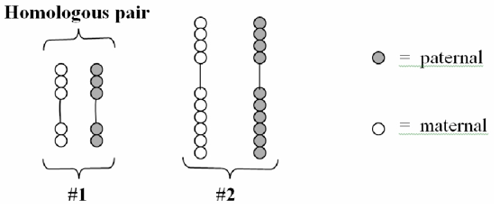
This is what your critter’s chromosomes look like in the unreplicated form. Note that there are four chromosomes here, or two homologous pairs. Each chromosome pair consists of a maternal and paternal version of the chromosome. The maternal and paternal versions are represented by respective bead color.
- Replicate your chromosomes! Make enough copies of each chromosome to represent both paternal and maternal chromosomes in a replicated form, as shown below. Note that the sister chromatids are identical in color. Be sure you can identify the sister chromatids, chromosomes, and the difference between a replicated and non-replicated form.

- Using your maternal and paternal sets of replicated chromosomes and your notes as a reference, practice the process of mitosis until you are very comfortable with it. Each person in the group should practice the entire process.
Think about It
Draw your bead chromosomes in each stage of mitosis. Label each stage. Note: You do not need to draw every single bead . . . but be sure to accurately indicate the relative sizes and colors of each different chromosome pair.
Do NOT proceed until you are comfortable with this! When your entire group is ready, let your instructor know. He or she will choose a group member to walk him or her through your simulation. If it is done correctly, you may move on to the next part.
Part 3: Microscopic Mitosis
In this part of the lab, you will examine 2 different slides:
- A cross section of an onion root tip, where cell growth (and consequently mitosis) happens at a rapid rate.
- Blastula of a whitefish. The blastula is a distinct stage during embryonic development when a fertilized egg forms a hollow ball of cells. During embryonic development, cells are dividing quickly and we are more likely to be able to see the varying stages of mitosis.
Materials
- Alium slide
- Whitefish blastula slide
- Microscope
Procedure
You must have your own microscope for this lab!
- Using correct microscope procedure, observe an onion root tip under high power (400X).
- Locate the region of active cell division, known as the root apical meristem, which is about 1 mm behind the actual tip of the root.
- Identify and draw a cell in each of the four stages of mitosis in the onion slide. Then draw cells in cytokinesis and interphase as well.
- Observe the prepared slide of a whitefish blastula under high power (400X).
- Identify and draw a cell in each of the four stages of mitosis in the whitefish blastula slide. Then draw cells in cytokinesis and interphase as well.
Part 4: Estimating Relative Time Spent in Each Stage of Mitosis
If you froze time and took a snapshot of a group of cells in a living organism, you could estimate the relative amount of time a cell spends in each stage of the cell cycle simply by counting the number of cells in each stage. For example, if there are 100 cells in your view and 90 of them are in prophase, you can assume that the cells spend most of the time in prophase.
In this part, you will practice identifying cells in the various stages of mitosis, and then you will estimate the relative amount of time a cell spends in each stage.
Materials
- Alium slide
- Microscope
Procedure
- Return to the slide of the onion root tip. Using correct microscope procedure, observe an onion root tip under high power (400X).
- Choose ONE view and then carefully COUNT the number of cells in each stage of the cell cycle. Feel free to estimate the total number of cells in each stage.
Data
| Interphase | Prophase | Metaphase | Anaphase | Telophase | |
|---|---|---|---|---|---|
| Number of cells in each phase: | |||||
| Percent of cells in each stage (see Equation 1) | |||||
| Estimated time a cell spends in each stage (see Equation 2) |
Equation 1: [latex]\displaystyle\text{percent of cells in state}=\frac{\text{cells in stage}}{\text{total cells}}\times{100}[/latex]
Equation 2: [latex]\displaystyle\text{Time spent in stage}=\text{(fraction of cells in stage)}\times(24\text{h})\times\bigg(\frac{1440\text{ min}}{24\text{ h}}\bigg)[/latex]
Think about It
Are there any problems with this estimate? How could you make this exercise more effective?
Part 5: Mitosis Bingo
Fill in the boxes with the names of the different stages of Mitosis. You will use the same stage multiple times. Then watch as different stages of mitosis are shown on the screen. Every time you see a stage on your card, cover that spot with a marker. You must cover all the spots on a card to win! (Printable version here.)

Lab Questions
- Describe how cytokinesis is different in plant cells and animal cells.
- Make an estimate of how long (relatively speaking) a cell stays in each stage of mitosis (not including interphase). How did you determine your estimates?
- Can you think of any reasons why cells contain a genetic program that tells the cell to commit suicide (apoptosis)? Give reasons why this would be.
- Is the G0 phase a truly resting phase? If the cells in G0 are not truly resting, why do you think we use the term resting to describe the state of these cells? [Hint: think of your nerve cells and muscle cells.]
Candela Citations
- Biology Labs. Authored by: Wendy Riggs. Provided by: College of the Redwoods. Located at: http://www.redwoods.edu. License: CC BY: Attribution
- Cell Cycle Checkpoint Images. Provided by: OpenStax. Located at: http://cnx.org/contents/b3c1e1d2-839c-42b0-a314-e119a8aafbdd@8.55:28/Concepts-of-Biology. Project: Concepts of Biology. License: CC BY: Attribution
- Derived from Mitosis Cells Sequence. Authored by: LadyofHats. Located at: https://commons.wikimedia.org/wiki/File:Mitosis_cells_sequence.svg. License: Public Domain: No Known Copyright