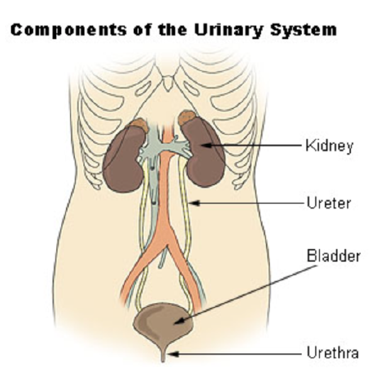
The actual human urinary system, also known as the renal system, is shown in the drawing below. The system consists of the kidneys, ureters, bladder, and urethra, which is the only structure not visible in the sculpture above. The main function of the urinary system is to eliminate the waste products of metabolism from the body by forming and excreting urine. Between 1 and 2 liters of urine are normally produced every day in a healthy individual.
Organs of the Urinary System
The urinary system is all about urine. It includes organs that form urine and also those that transport, store, or excrete urine.
Kidneys
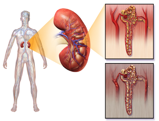
Urine is formed by the kidneys, which filter many substances out of blood, allow the blood to reabsorb needed materials, and use the remaining materials to form urine. The human body normally has two paired kidneys, although it is possible to get by quite well with just one kidney. As shown in the figure below, each kidney is well supplied with blood vessels by a major artery and vein. Blood to be filtered enters the kidney through the renal artery, and the filtered blood leaves the kidney through the renal vein. The kidney itself is wrapped in a fibrous capsule and consists of a thin outer layer called the cortex and a thicker inner layer called the medulla.

Blood is filtered and urine is formed by tiny functional units called nephrons. Each kidney contains at least a million nephrons, and each nephron spans the cortex and medulla layers of the kidney. After urine forms in the nephrons, it flows through a system of converging collecting ducts. The collecting ducts join together to form minor calyces, or chambers, which in turn join together to form major calyces (see the diagram above). Ultimately, the major calyces join the renal pelvis, which is the funnel-like end of the ureter where it enters the kidney.
Ureters, Bladder, Urethra
After urine forms in the kidneys, it is transported through the ureters (one per kidney) to the sac-like bladder, which stores the urine until urination. During urination, the urine is released from the bladder and transported by the urethra to be excreted outside the body through the external urethral opening.
Functions of the Urinary System
Waste products removed from the body with the formation and elimination of urine include many water-soluble metabolic products. The main waste products are urea, a by-product of protein catabolism, and uric acid, a by-product of nucleic acid catabolism. Excess water and mineral ions are also eliminated in urine.
Besides the elimination of waste products such as these, the urinary system has several other vital functions. These include:
- maintaining homeostasis of mineral ions in extracellular fluid. These ions are either excreted in urine or returned to the blood as needed to maintain the proper balance.
- regulating the acid-base balance in the body. For example, when pH is too low (blood is too acidic), the kidneys excrete less bicarbonate (which is basic) in the urine. When pH is too high (blood is too basic), the opposite occurs and more bicarbonate is excreted in the urine.
- controlling the volume of extracellular fluids, including the blood, which helps maintain blood pressure. The kidneys control fluid volume and blood pressure by excreting more or less salt and water in urine.
Control of the Urinary System
The formation of urine must be closely regulated to maintain body-wide homeostasis. Several endocrine hormones help control this function of the urinary system, including antidiuretic hormone, parathyroid hormone, and aldosterone.
- Antidiuretic hormone, also called vasopressin, is secreted by the hypothalamus. One of its main roles is conserving body water. It is released when the body is dehydrated and causes the kidneys to excrete less water in urine.
- Parathyroid hormone is secreted by the parathyroid glands. It works to regulate the balance of mineral ions in the body through its effects on several organs, including the kidneys. Parathyroid hormone stimulates the kidneys to excrete less calcium and more phosphorus in the urine.
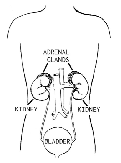
- Aldosterone is secreted by the cortex of the adrenal glands, which rest atop the kidneys, as shown in the figure below. It plays a central role in regulating blood pressure through its effects on the kidneys. It causes the kidneys to excrete less sodium and water in urine.
Once urine forms, it is excreted from the body in the process of urination. This process is controlled by both the autonomic and the somatic nervous systems. As the bladder fills with urine, it causes the autonomic nervous system to signal a muscle in the bladder wall to contract and the sphincter between the bladder and urethra to relax and open. This forces urine out of the bladder and through the urethra. Another sphincter at the distal end of the urethra is under voluntary control. When it relaxes under the influence of the somatic nervous system, it allows urine to leave the body through the external urethral opening.
Disorders of the Urinary System
Awareness Ribbon
Awareness ribbons are symbols meant to show support or raise consciousness for a cause. Different colors are associated with different issues, often relating to health problems. The first ribbon to gain familiarity for a health issue was the red ribbon for HIV/AIDS, created in 1991. The pink ribbon for breast cancer awareness is probably the best known today. Do you know what a green ribbon like the one pictured below represents? Among several other health problems, a green ribbon is meant to show support or raise awareness for kidney disorders.

The kidneys play such vital roles in eliminating wastes and toxins and maintaining body-wide homeostasis that disorders of the kidneys may be life-threatening. The gradual loss of normal kidney function commonly occurs with a number of disorders, including diabetes mellitus and high blood pressure. Other disorders of the kidneys are caused by faulty genes that are inherited. Loss of kidney function may eventually progress to kidney failure.
Diabetic Nephropathy
Diabetic nephropathy is a progressive kidney disease caused by damage to the capillaries in the glomeruli of the kidneys due to long-standing diabetes mellitus (see Figure 19.5.219.5.2). It is not fully understood how diabetes leads to damage of glomerular capillaries, but it is thought that high levels of glucose in the blood are involved. In people with diabetes, nephropathy is more likely if their blood glucose is poorly controlled. Having high blood pressure, a history of cigarette smoking and a family history of kidney problems are additional risk factors. Diabetic nephropathy often has no symptoms at first. In fact, it may take up to a decade after kidney damage begins for symptoms to appear. When they do appear, they typically include severe tiredness, headaches, nausea, frequent urination, and itchy skin.
Proteins are large molecules that usually are not filtered out of blood in the glomeruli. When the glomerular capillaries are damaged, it allows proteins such as albumin to leak into the filtrate from the blood. As a result, albumin ends up being excreted in the urine. Finding a high level of albumin in the urine is one indicator of diabetic nephropathy and helps to diagnose the disorder. Drugs may be prescribed to reduce protein levels in the urine. Controlling high blood sugar levels and hypertension (high blood pressure) is also important to help slow kidney damage, as is a reduction of sodium intake.
Polycystic Kidney Disease
Polycystic kidney disease (PKD) is a genetic disorder in which multiple abnormal cysts develop and grow in the kidneys. The photo below shows a pair of kidneys that are riddled with cysts from PKD. In people who inherit PKD, the cysts may start to form at any point in life from infancy through adulthood. Typically, both kidneys are affected. Symptoms of the disorder may include high blood pressure, headaches, abdominal pain, blood in the urine, and excessive urination.
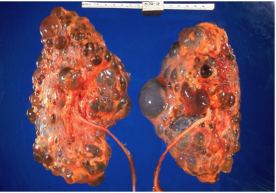
There are two types of PKD. The more common type is caused by an autosomal dominant allele, and the less common type is caused by an autosomal recessive allele. Both types collectively make PKD one of the most common hereditary diseases in the United States, affecting more than half a million people. There is little or no difference in the rate of occurrence of PKD between genders or ethnic groups. There is no known cure for this disease other than a kidney transplant.
Kidney Failure
Both diabetic nephropathy and PKD may lead to kidney (or renal) failure (classified as end-stage kidney disease), in which the kidneys are no longer able to adequately filter metabolic wastes from the blood. Long-term, uncontrolled high blood pressure is another common cause of kidney failure. Symptoms of kidney failure may include nausea, more or less frequent urination, blood in the urine, muscle cramps, anemia, swelling of the extremities, and shortness of breath due to the accumulation of fluid in the lungs. If kidney function drops below the level needed to sustain life, then the only treatment option is kidney transplantation or some means of artificial filtration of the blood, such as by hemodialysis.
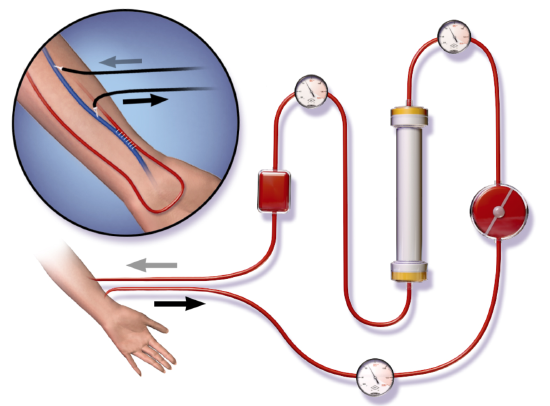
Hemodialysis is a medical procedure in which blood is filtered externally through a machine. You can see how it works in the simplified diagram below. During dialysis, waste products such as urea as well as excess water are removed from the patient’s blood before the blood is returned to the patient. Hemodialysis is typically done on an outpatient basis in a hospital or special dialysis clinic. Less frequently, it is done in the patient’s home. Depending on the patient’s size, among other factors, the blood is filtered for 3 to 4 hours about 3 times a week. Because the treatment is needed so frequently, hemodialysis is one of the most common procedures performed in U.S. hospitals.
Kidney Stones
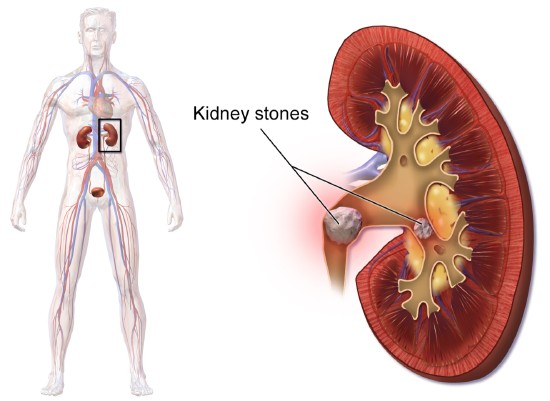
A kidney stone, also known as a renal calculus, is a solid crystal that forms in a kidney from minerals in urine (see Figure 19.5.519.5.5). The majority of kidney stones consist of crystals of calcium salts. Kidney stones typically leave the body in the urine stream. A small stone may pass through the ureters and other urinary tract organs without causing symptoms and go undetected. A larger stone may cause pain when it passes through the urinary tract. If a kidney stone grows large enough, it may block the ureter. Blockage of a ureter may cause a decrease in kidney function and damage to the kidney.
A kidney stone that causes pain is generally treated with pain medication until it passes through the urinary tract. A stone that causes a blockage may be treated with lithotripsy. This is a medical procedure in which high-intensity ultrasound pulses are applied externally to cause fragmentation of the stone into pieces small enough to pass easily through the urinary tract. Although lithotripsy is noninvasive, it can cause damage to the kidneys. An alternative treatment for a stone that blocks urine flow is to insert a stent into the ureter to expand it and allow both urine and the stone to pass. In some cases, surgery may be required to physically remove a large stone from the ureter.
A combination of lifestyle and genetic factors seem to predispose certain people to develop kidney stones. Risk factors include high consumption of cola soft drinks, eating a diet high in animal protein, being overweight, and not drinking enough fluids. Preventive measures are obvious. They include limiting cola consumption, eating less animal protein, losing weight, and increasing fluid intake.
Other Urinary System Disorders
Although disorders of the kidneys are generally the most serious urinary system disorders, problems that affect other organs of the urinary tract are generally more common. They include bladder infections and urinary incontinence.
Bladder Infection
A bladder infection, also called cystitis, is a very common type of urinary tract infection in which the urinary bladder becomes infected by bacteria (typically Escherichia coli), rarely by fungi. Symptoms of bladder infections may include pain with urination, frequent urination, and feeling the need to urinate despite having an empty bladder. In some cases, there may be blood in the urine. A much less common type of urinary tract infection is pyelonephritis, in which the kidney becomes infected. If a kidney infection occurs, it is generally because of an untreated bladder infection. Bladder infections are treated mainly with antibiotics.
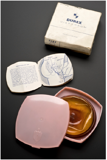
Risk factors for urinary bladder infections include sexual intercourse (especially when spermicide or a diaphragm, as shown below, is used for contraception), diabetes, obesity, and most importantly, female sex. Bladder infections are four times more common in women than in men. In fact, in women, they are the most common type of bacterial infection, and as many as 1 in 10 women has a bladder infection in any given year. Female anatomy explains the sex difference in the incidence of bladder infections. The urethra is much shorter and closer to the anus in females than in males, so contamination of the urethra and then the bladder with GI tract bacteria is more likely in females than in males. Once the bacteria reach the bladder, they can attach to the bladder wall and form a biofilm that resists the body’s immune response.
Urinary Incontinence
Urinary incontinence is a chronic problem of uncontrolled leakage of urine. It is very common, especially at older ages and especially in women. Sometimes urinary incontinence is a sign of another health problem, such as diabetes or obesity. Regardless of the underlying cause, the symptoms of urinary incontinence alone may have a large impact on the quality of life, frequently causing inconvenience, embarrassment, and distress.
In a person with male anatomy, urinary incontinence is most commonly caused by an enlarged prostate gland or treatment for prostate cancer. In genetically female individuals, there are two common types of urinary incontinence with different causes: stress incontinence and urge incontinence.
- Stress urinary incontinence is caused by loss of support of the urethra, usually due to stretching of pelvic floor muscles during childbirth. It is characterized by leakage of small amounts of urine with activities that increase abdominal pressure, such as coughing, sneezing, or lifting. Treatment of stress urinary incontinence may include Kegel exercises to strengthen the pelvic muscles (see Explore More below). More serious cases may call for surgery to improve support for the bladder.
- Urge urinary incontinence (commonly called “overactive bladder”) is caused by uncontrolled contractions of the detrusor muscle in the wall of the bladder. This causes the bladder to empty unexpectedly. Urge incontinence is characterized by leakage of large amounts of urine in association with an insufficient warning to get to the bathroom in time. Treatment of urge incontinence may include taking medication to relax the detrusor muscle.
Candela Citations
- Introduction to the Urinary System. Authored by: Contributed by Suzanne Wakim & Mandeep Grewal Professors (Cell Molecular Biology & Plant Science) at Butte College. Located at: https://bio.libretexts.org/Bookshelves/Human_Biology/Book%3A_Human_Biology_(Wakim_and_Grewal)/19%3A_Urinary_System/19.2%3A_Introduction_to_the_Urinary_System. Project: Book: Human Biology (Wakim & Grewal). License: CC BY-NC: Attribution-NonCommercial