What is Respiration?
Respiration is the life-sustaining process in which gases are exchanged between the body and the outside atmosphere. Specifically, oxygen moves from the outside air into the body; and water vapor, carbon dioxide, and other waste gases move from inside the body into the outside air. Respiration is carried out mainly by the respiratory system. It is important to note that respiration by the respiratory system is not the same process as cellular respiration that occurs inside cells, although the two processes are closely connected. Cellular respiration is the metabolic process in which cells obtain energy, usually by “burning” glucose in the presence of oxygen. When cellular respiration is aerobic, it uses oxygen and releases carbon dioxide as a waste product. Respiration by the respiratory system supplies the oxygen needed by cells for aerobic cellular respiration and removes the carbon dioxide produced by cells during cellular respiration.
Respiration by the respiratory system actually involves two subsidiary processes. One process is ventilation or breathing. This is the physical process of conducting air to and from the lungs. The other process is gas exchange. This is the biochemical process in which oxygen diffuses out of the air and into the blood while carbon dioxide and other waste gases diffuse out of the blood and into the air. All of the organs of the respiratory system are involved in breathing, but only the lungs are involved in gas exchange.
Respiratory Organs
The organs of the respiratory system form a continuous system of passages called the respiratory tract, through which air flows into and out of the body. The respiratory tract has two major divisions: the upper respiratory tract and the lower respiratory tract. The organs in each division are shown in the figure below. In addition to these organs, certain muscles of the thorax (the body cavity that fills the chest) are also involved in respiration by enabling breathing. Most important is a large muscle called the diaphragm, which lies below the lungs and separates the thorax from the abdomen. Smaller muscles between the ribs also play a role in breathing. You can learn more about breathing muscles in the concept of Breathing.
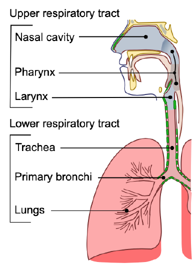
Upper Respiratory Tract
All of the organs and other structures of the upper respiratory tract are involved in conduction or the movement of air into and out of the body. Upper respiratory tract organs provide a route for air to move between the outside atmosphere and the lungs. They also clean, humidity, and warm the incoming air. However, no gas exchange occurs in these organs.
Nasal Cavity
The nasal cavity is a large, air-filled space in the skull above and behind the nose in the middle of the face. It is a continuation of the two nostrils. As inhaled air flows through the nasal cavity, it is warmed and humidified. Hairs in the nose help trap larger foreign particles in the air before they go deeper into the respiratory tract. In addition to its respiratory functions, the nasal cavity also contains chemoreceptors that are needed for the sense of smell and that contribute importantly to the sense of taste.
Pharynx
The pharynx is a tube-like structure that connects the nasal cavity and the back of the mouth to other structures lower in the throat, including the larynx. The pharynx has dual functions: both air and food (or other swallowed substances) pass through it, so it is part of both the respiratory and the digestive systems. Air passes from the nasal cavity through the pharynx to the larynx (as well as in the opposite direction). Food passes from the mouth through the pharynx to the esophagus.
Larynx
The larynx connects the pharynx and trachea and helps to conduct air through the respiratory tract. The larynx is also called the voice box because it contains the vocal cords, which vibrate when air flows over them, thereby producing sound. You can see the vocal cords in the larynx in the figure below. Certain muscles in the larynx move the vocal cords apart to allow breathing. Other muscles in the larynx move the vocal cords together to allow the production of vocal sounds. The latter muscles also control the pitch of sounds and help control their volume.
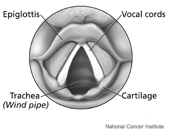
A very important function of the larynx is protecting the trachea from aspirated food. When swallowing occurs, the backward motion of the tongue forces a flap called the epiglottis to close over the entrance to the larynx. (You can see the epiglottis in the figure above.) This prevents swallowed material from entering the larynx and moving deeper into the respiratory tract. If swallowed material does start to enter the larynx, it irritates the larynx and stimulates a strong cough reflex. This generally expels the material out of the larynx and into the throat.
Lower Respiratory Tract
The trachea and other passages of the lower respiratory tract conduct air between the upper respiratory tract and the lungs. These passages form an inverted tree-like shape (Figure 16.2.416.2.4), with repeated branching as they move deeper into the lungs. All told, there are an astonishing 1,500 miles of airways conducting air through the human respiratory tract! It is only in the lungs, however, that gas exchange occurs between the air and the bloodstream.
Trachea
The trachea, or windpipe, is the widest passageway in the respiratory tract. It is about 2.5 cm (1 in.) wide and 10-15 cm (4-6 in.) long. It is formed by rings of cartilage, which make it relatively strong and resilient. The trachea connects the larynx to the lungs for the passage of air through the respiratory trac—thee trachea branches at the bottom to form two bronchial tubes.
Bronchi and Bronchioles
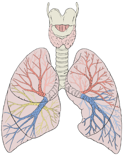
There are two main bronchial tubes, or bronchi (singular, bronchus), called the right and left bronchi. The bronchi carry air between the trachea and lungs. Each bronchus branches into smaller, secondary bronchi; and secondary bronchi branch into still smaller tertiary bronchi. The smallest bronchi branch into very small tubules called bronchioles. The tiniest bronchioles end in alveolar ducts, which terminate in clusters of minuscule air sacs, called alveoli (singular, alveolus), in the lungs.
Lungs
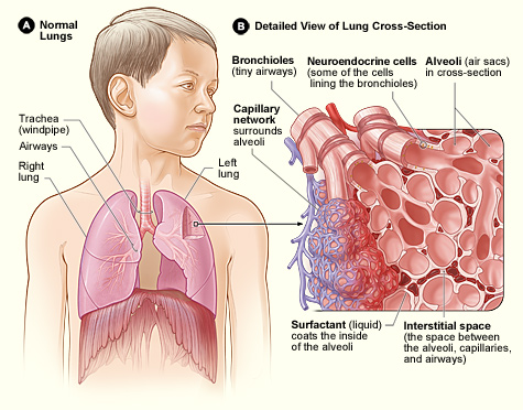

The lungs are the largest organs of the respiratory tract. They are suspended within the pleural cavity of the thorax. The lungs are surrounded by two thin membranes called pleura, which secrete a fluid that allows the lungs to move freely within the pleural cavity. This is necessary so the lungs can expand and contract during breathing. In Figure 16.2.516.2.5, you can see that each of the two lungs is divided into sections. These are called lobes, and they are separated from each other by connective tissues. The right lung is larger and contains three lobes. The left lung is smaller and contains only two lobes. The smaller left lung allows room for the heart, which is just left of the center of the chest.
Lung tissue consists mainly of alveoli (Figure 16.2.616.2.6). These tiny air sacs are the functional units of the lungs where gas exchange takes place. The two lungs may contain as many as 700 million alveoli, providing a huge total surface area for gas exchange to take place. In fact, alveoli in the two lungs provide as much surface area as half a tennis court! Each time you breathe in, the alveoli fill with air, making the lungs expand. Oxygen in the air inside the alveoli is absorbed by the blood in the mesh-like network of tiny capillaries that surrounds each alveolus. The blood in these capillaries also releases carbon dioxide into the air inside the alveoli. Each time you breathe out, air leaves the alveoli and rushes into the outside atmosphere, carrying waste gases with it.
The lungs receive blood from two major sources. They receive deoxygenated blood from the heart. This blood absorbs oxygen in the lungs and carries it back to the heart to be pumped to cells throughout the body. The lungs also receive oxygenated blood from the heart that provides oxygen to the cells of the lungs for cellular respiration.
Protecting the Respiratory System
You may be able to survive for weeks without food and for days without water, but you can survive without oxygen for only a matter of minutes except under exceptional circumstances. Therefore, protecting the respiratory system is vital. That’s why making sure a patient has an open airway is the first step in treating many medical emergencies. Fortunately, the respiratory system is well protected by the ribcage of the skeletal system. However, the extensive surface area of the respiratory system is directly exposed to the outside world and all its potential dangers in inhaled air. Therefore, it should come as no surprise that the respiratory system has a variety of ways to protect itself from harmful substances such as dust and pathogens in the air.
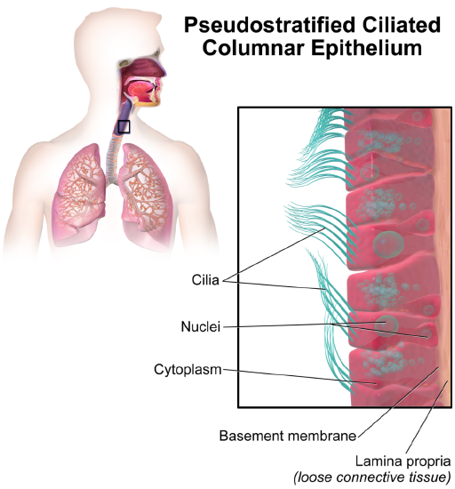
The main way the respiratory system protects itself is called the mucociliary escalator. From the nose through the bronchi, the respiratory tract is covered in the epithelium that contains mucus-secreting goblet cells. The mucus traps particles and pathogens in the incoming air. The epithelium of the respiratory tract is also covered with tiny cell projections called cilia (singular, cilium), as shown in the figure below. The cilia constantly move in a sweeping motion upward toward the throat, moving the mucus and trapped particles and pathogens away from the lungs and toward the outside of the body.
What happens to the material that moves up the mucociliary escalator to the throat? It is generally removed from the respiratory tract by clearing the throat or coughing. Coughing is a largely involuntary response of the respiratory system that occurs when nerves lining the airways are irritated. The response causes air to be expelled forcefully from the trachea, helping to remove mucus and any debris it contains (called phlegm) from the upper respiratory tract to the mouth. The phlegm may spit out (expectorated), or it may be swallowed and destroyed by stomach acids.
Sneezing is a similar involuntary response that occurs when nerves lining the nasal passage are irritated. It results in forceful expulsion of air from the mouth, which sprays millions of tiny droplets of mucus and other debris out of the mouth and into the air, as shown in the photo below. This explains why it is so important to sneeze into a sleeve rather than the air to help prevent the transmission of respiratory pathogens.
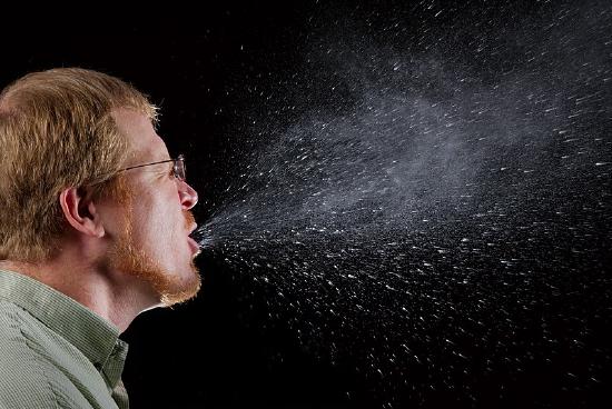
Figure 16.2.816.2.8: Sneezing results in tiny particles from the mouth being forcefully ejected into the air.
How the Respiratory System Works with Other Organ Systems
The amount of oxygen and carbon dioxide in the blood must be maintained within a limited range for the survival of the organism. Cells cannot survive for long without oxygen, and if there is too much carbon dioxide in the blood, the blood becomes dangerously acidic (pH is too low). Conversely, if there is too little carbon dioxide in the blood, the blood becomes too basic (pH is too high). The respiratory system works hand-in-hand with the nervous and cardiovascular systems to maintain homeostasis in blood gases and pH.
It is the level of carbon dioxide rather than the level of oxygen that is most closely monitored to maintain blood gas and pH homeostasis. The level of carbon dioxide in the blood is detected by cells in the brain, which speed up or slow down the rate of breathing through the autonomic nervous system as needed to bring the carbon dioxide level within the normal range. Faster breathing lowers the carbon dioxide level (and raises the oxygen level and pH); slower breathing has the opposite effects. In this way, the levels of carbon dioxide and oxygen, as well as pH, are maintained within normal limits.
The respiratory system also works closely with the cardiovascular system to maintain homeostasis. The respiratory system exchanges gases between the blood and the outside air, but it needs the cardiovascular system to carry them to and from body cells. Oxygen is absorbed by the blood in the lungs and then transported through a vast network of blood vessels to cells throughout the body where it is needed for aerobic cellular respiration. The same system absorbs carbon dioxide from cells and carries it to the respiratory system for removal from the body.
Feature: My Human Body
Choking due to a foreign object becoming lodged in the airway results in close to 200,000 deaths each year. For the sake of your own human body, as well as those of loved ones, you should be aware of choking risks, signs, and treatments.
Choking is the mechanical obstruction of the flow of air from the atmosphere into the lungs. It prevents breathing and may be partial or complete. Partial choking allows some though inadequate airflow into the lung—prolonged or complete choking results in asphyxia, or suffocation, which is potentially fatal.
Obstruction of the airway typically occurs in the pharynx or trachea. Young children are more prone to choking than are older people, in part because they often put small objects in their mouths and do not appreciate the risk of choking that they pose. Young children may choke on small toys or parts of toys or on household objects in addition to food. Foods that can adapt their shape to that of the pharynx, such as bananas and marshmallows, are especially dangerous and may cause choking in adults as well as children.
How can you tell if a loved one is choking? The person cannot speak or cry out or has great difficulty doing so. Breathing, if possible, is labored, producing gasping or wheezing. The person may desperately clutch at his or her throat or mouth. If breathing is not soon restored, the person’s face will start to turn blue from lack of oxygen. This will be followed by unconsciousness if oxygen deprivation continues beyond a few minutes.
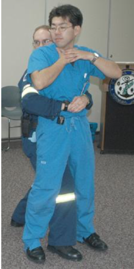
If an infant is choking, turning the baby upside down and slapping on the back may dislodge the obstructing object. To help an older person who is choking, first, encourage the person to cough. Give them a few hardback slaps to help force the lodged object out of the airway. If these steps fail, perform the Heimlich maneuver on the person. You can easily find instructional videos online to learn how to do it. If the Heimlich maneuver also fails, call for emergency medical care immediately.
Disorders of the Respiratory System
Asthma
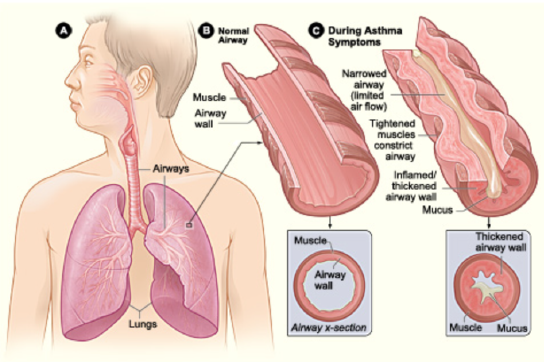
Asthma is a chronic inflammatory disease of the airways in the lungs, in which the airways periodically become inflamed. As you can see in Figure 16.4.216.4.2, this causes swelling and narrowing of the airways, often accompanied by excessive mucus production. Symptoms of asthma include difficulty breathing, coughing, wheezing, shortness of breath, and chest tightness. Some people with asthma rarely experience symptoms, and then usually only in response to certain triggers in the environment. Other people may have symptoms almost all of the time.
Asthma is thought to be caused by a combination of genetic and environmental factors. A person with a family history of asthma is more likely to develop the disease. Dozens of genes have been found to be associated with asthma, many of which are related to the immune system. Additional risk factors include obesity and sleep apnea (see the feature My Human Body below). Environmental factors trigger asthma attacks in people who have a genetic predisposition to the disease. Besides dust mite feces, triggers may include other allergens (such as pet dander, cockroaches, and mold), certain medications including aspirin, air pollution, and stress, among other possible factors. Symptoms tend to be worse at night and early in the morning. They may also worsen during upper respiratory tract infections, strenuous exercise, or when the airways are exposed to cold air.
There is no cure for asthma at present, but the symptoms of asthma attacks usually can be reversed with the use of inhaled medications called bronchodilators. These medications soothe the constricted air passages and help to re-expand them, making breathing easier. The medications usually start to take effect almost immediately. Other medications can be taken for long-term control of the disease. These medications help prevent asthma attacks from occurring. Corticosteroids are generally considered the most effective treatment for long-term control. Another way to prevent asthma attacks is by avoiding triggers whenever possible.
Pneumonia
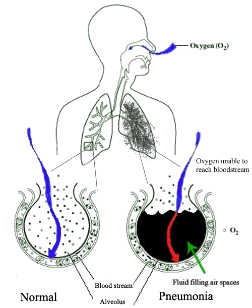
Another common inflammatory disease of the respiratory tract is pneumonia. In pneumonia, the inflammation affects primarily the alveoli, which are the tiny air sacs of the lungs. Inflammation causes some of the alveoli to become filled with fluid so that gas exchange cannot occur. This is illustrated in the figure below. Symptoms of pneumonia typically include coughing, chest pain, difficulty breathing, and fever.
Pneumonia often develops as a consequence of an upper respiratory tract infection such as the common cold or flu, especially in the very young and the elderly. It is usually caused by bacteria or viruses, although some cases may be caused by other microorganisms such as fungi. The majority of cases are caused by just a few pathogens, the most common being the bacterium Streptococcus pneumoniae. Pneumonia is more likely to develop in people who have other lung diseases such as asthma, a history of smoking, heart failure, or a weakened immune system.
Vaccines are available to prevent certain types of bacterial and viral pneumonia, including pneumonia caused by Streptococcus pneumoniae. Treatment of pneumonia depends on the cause. For example, if it is caused by bacteria, antibiotics are generally prescribed. In cases of severe pneumonia, hospitalization and supplemental oxygen may be required.
Chronic Obstructive Pulmonary Disease
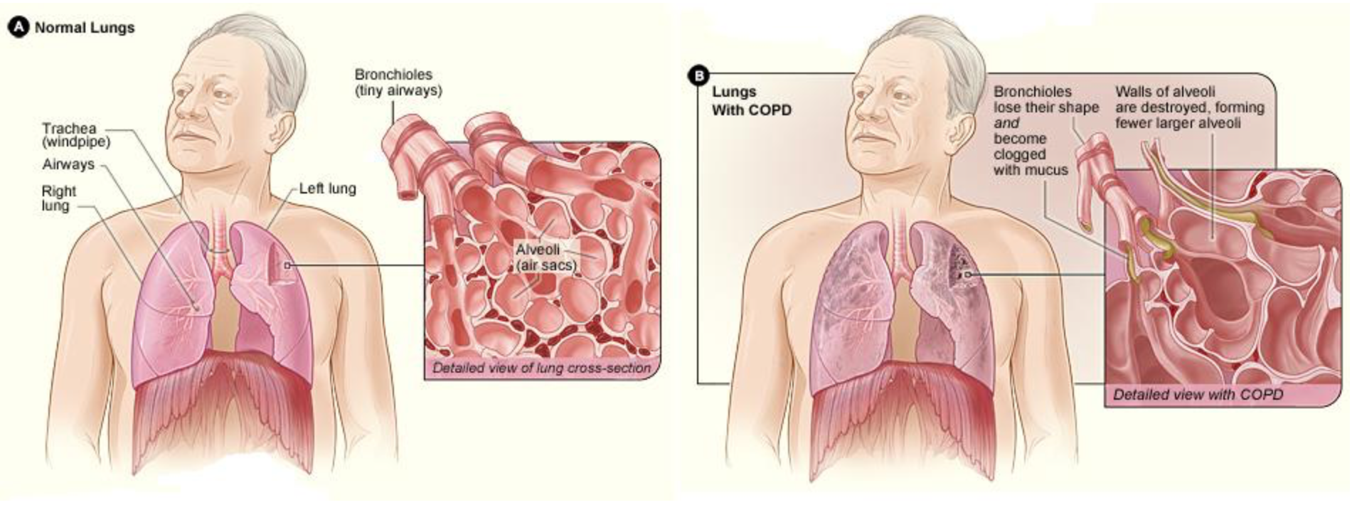
Chronic obstructive pulmonary disease (COPD) is a lung disease characterized by chronic poor airflow. The main symptoms include shortness of breath and a cough that produces phlegm. These symptoms are usually present for a long period of time and typically become worse over time. Eventually, walking up stairs and similar activities become difficult because of shortness of breath.
COPD formerly was referred to as chronic bronchitis or emphysema. Now, the term chronic bronchitis is used to refer to the symptoms of COPD, and the term emphysema is used to refer to the lung changes that occur with COPD. Some of these lung changes are shown in the figure below. They include a breakdown of connective tissues that reduces the number and elasticity of alveoli. As a result, the patient can no longer fully exhale air from the lungs, so air becomes trapped in the lungs. Gas exchange is hampered and may lead to low oxygen levels and too much carbon dioxide in the blood.
Tobacco smoking is the major cause of COPD, with a number of other factors such as air pollution and genetics playing smaller roles. Of people who are life-long smokers, about half will eventually develop COPD. Exposure to secondhand smoke in nonsmokers also increases the risk of COPD and accounts for about 20 percent of cases. Most cases of COPD could have been prevented by never smoking. In people who have already been diagnosed with COPD, cessation of smoking can slow down the rate at which COPD worsens. People with COPD may be treated with supplemental oxygen and inhaled bronchodilators. These treatments may reduce the symptoms but there is no cure for COPD except, in very severe cases, lung transplantation (see the Explore More video below).
Lung Cancer
Lung cancer is a malignant tumor characterized by uncontrolled cell growth in tissues of the lung. The tumor may arise directly from lung tissue (primary lung cancer) or as a result of metastasis from cancer in another part of the body (secondary lung cancer). Primary lung cancer may also metastasize and spread to other parts of the body. Lung cancer develops following genetic damage to DNA that affects the normal functions of the cell. As more damage accumulates, the risk of cancer increases. The most common symptoms of lung cancer include coughing (especially coughing up blood), wheezing, shortness of breath, chest pain, and weight loss.
The major cause of primary lung cancer is tobacco smoking, which accounts for about 85 percent of cases. Cigarette smoke contains numerous cancer-causing chemicals. Besides smoking, other potential causes of lung cancer include exposure to radon gas, asbestos, secondhand smoke, or other air pollutants. When tobacco smoking is combined with other risk factors such as exposure to radon or asbestos, the risk of lung cancer is heightened. People who have close biological relatives with lung cancer are also at increased risk of developing the disease.
Most cases of lung cancer cannot be cured. In many people, cancer has already spread beyond the original site by the time they have symptoms and seek medical attention. About 10 percent of people with lung cancer do not have symptoms when they are diagnosed, and the cancers are found when they have a chest X-ray for another problem. In part because of its typically late diagnosis, lung cancer is the most common cause of cancer-related death in men and the second most common cause in women (after breast cancer). Common treatments for lung cancer include surgical removal of the tumor, radiation therapy, chemotherapy, or some combination of these three types of treatment.
Feature: My Human Body
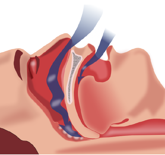
Do you — or someone you love — snore? Snoring may be more than just an annoyance. It may also be a sign of a potentially dangerous and common disorder known as sleep apnea. Sleep apnea is characterized by pauses in breathing that occur most often because of physical blockage to airflow during sleep. When breathing is paused, carbon dioxide builds up in the bloodstream. The higher-than-normal level of carbon dioxide in the blood causes the respiratory centers in the brain to wake the person enough to start breathing normally. This reduces the carbon dioxide level, and the person falls back asleep. This occurs repeatedly throughout the night, causing serious disruption in sleep. Most people with sleep apnea are unaware that they have the disorder because they don’t awake fully enough to remember the repeated awakenings throughout the night. Instead, sleep apnea is more commonly recognized by other people who witness the episodes.
The figure below shows how sleep apnea typically occurs. The muscle tone of the body normally relaxes during sleep, allowing the soft tissues in the throat to collapse and block the airway. The relaxation of muscles may be exacerbated by the use of alcohol, tranquilizers, or muscle relaxants. The risk of sleep apnea is greater in people who are overweight, smoke tobacco, or have diabetes. The disorder is also more likely to occur in older people and males. Common symptoms of sleep apnea include loud snoring, restless sleep, and daytime sleepiness and fatigue. Daytime sleepiness, in turn, increases the risk of driving and work-related accidents. Continued sleep deprivation may cause moodiness and belligerence. Lack of adequate oxygen to the body because of sleep apnea may also lead to other health problems including fatty liver diseases and high blood pressure. Symptoms of sleep apnea may be present for years or even decades until (and if) a diagnosis is finally made.
Treatment of sleep apnea may include avoiding alcohol, quitting smoking, or losing weight. Elevating the upper body during sleep or sleeping on one’s side may help prevent airway collapse in many people with sleep apnea. Another type of treatment is the use of an oral device during sleep that shifts the lower jaw forward to help keep the airway open. The most common treatment for moderate to severe sleep apnea is the use during sleep of CPAP (continuous positive airway pressure), which keeps the airway open by means of pressurized air. In this treatment, the person typically wears a plastic facial mask that is connected by a flexible tube to a small bedside CPAP machine. Although CPAP is effective, long-term compliance is often poor because patients find the mask uncomfortable or they experience unpleasant side effects such as dry mouth and nose. A more extreme form of treatment is surgery to remove some of the tissues — such as the tonsils or part of the soft palate — that tend to collapse and block the airway in people with sleep apnea.
watch
Check out this video to learn about how the immune system can overreact to cause asthma:
Candela Citations
- Structure and Function of the Respiratory System. Authored by: Contributed by Suzanne Wakim & Mandeep Grewal Professors (Cell Molecular Biology & Plant Science) at Butte College. Located at: https://bio.libretexts.org/Bookshelves/Human_Biology/Book%3A_Human_Biology_(Wakim_and_Grewal)/16%3A_Respiratory_System/16.2%3A_Structure_and_Function_of_the_Respiratory_System. Project: Book: Human Biology (Wakim & Grewal). License: CC BY-NC: Attribution-NonCommercial